Human Platelet Extract: Some People Want You to Apply Blood Clotting, Cancer-Causing Factors to Your Face

In the land of where some people will do nearly anything for money, a company is selling “human platelet extract” as a topical cosmetic product for daily use on normal skin. Can you say, “Gee, let’s clot our skin’s blood supply, induce inflammation, and induce cancer at the same time?” Yes, extended use of a platelet extract product is likely carcinogenic (Carr et al, 2014; Sabrkhany et al, 2021).  To be clear, platelet lysate (extract) triggers an inflammatory response in stem cells in the skin and induces the secretion of factors maintaining immune cells (macrophages) in a proinflammatory state thus enhancing inflammation (Ulivi et al, 2014), and long term inflammation is dangerous.

First, platelet extract does not contain exosomes as some are claiming. The company Plated uses the term “Renewosome”” to fool people. It’s marketing hype that the hysterical mass media has been repeating. They even put a trademark after this silly name. Lol. Platelet-derived exosomes (PLT-Exos) are the main subtype of extracellular vesicles secreted by platelets, which carry proteins, nucleotides, lipids, and other substances to acceptor cells, playing an important role in intercellular communication.” Notice the term “secreted.” Exosomes are actively secreted from living platelets, and an extraction process of platelets will not yield any exosomes. None. I introduced the concept of and actual products containing exosomes to skin care over a decade ago. Many people criticized me at the time, and never bothered to read my peer-reviewed work on the subject, including my scientific book on the subject. But over a decade later following my introduction of exosomes to skin care, the word is out and people are making false claims to have exosomes in their products. Again, exosomes must be released (secreted) and cannot be collected through cellular extraction processes.

BTW, if you want to buy some human platelet extract (lysate is the scientific term for extract), here’s the source:

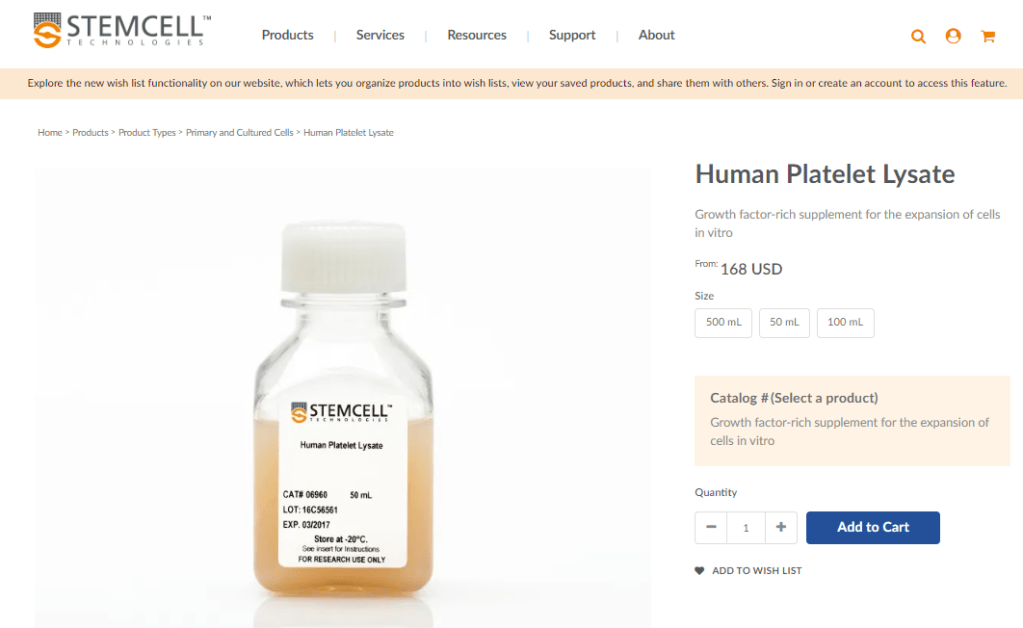
You can also purchase the platelet extract without fibrinogen. The company has removed the fibrinogen to reduce clotting because fibrinogen is one of the clotting factors.

So, what are platelets? Platelets are anucleated red blood cells that circulate in blood. They are small because they’re anucleated and can therefore squeeze into small places. Platelets are called into action when a wound occurs and blood is spewing. Their job is to close the wound fast by clotting to stop blood leakage, and by inducing high rates of cellular proliferation and inducing an inflammatory response to fight infection. Thus, quoting from Scherlinger et al (2023) in a Nature review article, “platelets produce soluble factors and directly interact with immune cells, thereby promoting an inflammatory phenotype. Furthermore, platelets participate in tissue injury and promote abnormal tissue healing, leading to fibrosis.” Inflammation, fibrosis, abnormal tissue healing with an abnormal matrix, and proliferation are hallmarks of cancer. Tumors are wounds that don’t heal, and applying platelet extract on your face mimics a wound that doesn’t heal. Platelets don’t live long in humans, about 7-10 days, nor would you want them to. They’ve evolved to flux into an area rapidly, secrete all of their inflammatory, clotting, and proliferative factors, close the wound, and then die before before they cause too much damage. If the platelets didn’t die, but instead stayed around for a long time secreting their inflammatory, clotting, and proliferative factors, it would be similar to applying platelet extract to your skin on daily basis. Chronic inflammation, clotting and fibrosis with tumorigenesis would result.

If we consider platelet rich plasma (PRP), a less concentrated form of platelet extract, an inflammatory response in fibroblasts is induced that leads to the formation of ROS (reactive oxygen species) and activation of oxidative stress pathways. It does not promote regeneration. Recent studies have found, “Treatment with PRP increased reticular dermis thickness with a fibrotic aspect. In the long term, the presence of inflammation and microangiopathy caused by PRP injection could lead to trophic alteration of the skin and the precocious aging process.” In other words, platelets cause fibrosis and advanced aging of the skin. Why someone would want to use platelet extract on their skin is beyond me.

According to epidemiologists, “A growing body of laboratory research has shown the direct involvement of platelets with cancer.: Cancer follows a high platelet count. Signaling by platelet-derived growth factors (PDGFs) and their receptors (PDGFRs) is commonly observed in epithelial cancers, where it triggers stromal recruitment and may be involved in epithelial–mesenchymal transition, thereby affecting tumor growth, angiogenesis, invasion, and metastasis. In other words, the extract of platelets will be high in PDGF and when applied daily will enhance the probability of cancer.

Recent studies have found that over-active PDGF signaling is implicated in several types of human malignancies, in one way by promoting proliferation, survival and invasion of tumour cells directly, and in another way by changing the tumor stroma (matrix) in a manner that promotes tumorigenesis. If we look at a product called Regranex, which is a topical product containing 0.01% PDGF, the label includes a warning, “Malignancies distant from the site of application have occurred in REGRANEX users in a clinical study and in postmarketing use.” Thus, using a platelet extract, loaded with PDGF, on a daily basis may be asking for trouble, specifically tumorigenesis.

Let’s look at the ingredients of the platelet extract product used in a “clinical study” of the platelet extract product (from their website):

“purified water, glycerin, pentylene glycol, trideceth-9, panthenol, human platelet extract™, PEG-5 isononanoate, polyacrylate crosspolymer-6, hyaluronic acid, caprylyl glycol, 1,2-hexanediol, hydrolyzed gelatin, arginine, silanetriol, saccharide isomerate, menthyl lactate, carnosine, citric acid, sodium citrate.”

Question – If the “human platelet extract” is so good, why do they need to include all of those other actives?

Answer- because the platelet extract doesn’t work well on its own. At NeoGenesis, we don’t have to add anything to our S2RM adult stem cell-based technology containing exosomes because it really works.

Let’s look at a study published by the company selling platelet extract. The study is:

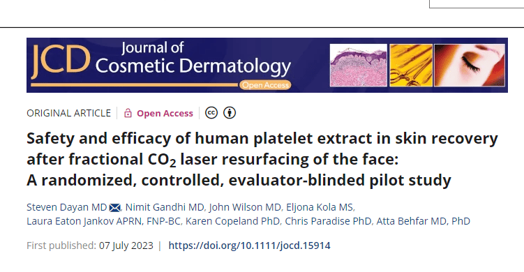
This study is for short term results in treated skin, not long term results in normal skin. The study suffers from a poor experimental design, conflicts of interest, and the results are underwhelming,

First, the study was not conducted at Mayo Clinic as some people have said in social media. Rather the study was performed for payment from the company to a plastic surgeon named Steve Dyan, and the authors included those employed by the company. So Steve Dyan put his name on the paper for money – this is common, and is called ghostwriting. This is where a physician puts their name on a paper when others have done the work. He was paid to put his name on this published paper. This is a conflict of interest, and is one of the reasons why most medical research and clinical trials cannot be believed.

Look at the study design, and find the problem with the design:

Treatment Group – Before Procedure:   “A 7-day pre-procedure facial skincare regimen for subjects randomized to the HPE treatment group included Cetaphil cleanser (or equivalent) twice daily morning and evening, with application of HPE once daily, and EltaMD (Colgate-Palmolive, New York, NY) UV Daily Broad-Spectrum SPF 40 (or equivalent) in the morning, with reapplication throughout the day as needed.”
Treatment Group – Post-Procedure: 

 “Post-procedure skincare (until the skin was fully healed at 7–10 days) in the treatment group included application of HPE three times daily (morning, mid-day, and before bedtime) followed by Vanicream (PSI, Rochester, MN) Moisturizing Ointment as needed for dryness, applied 15 min after HPE CALM. Post-healing when were they [sic] determined “healed” skincare in the HPE treatment group included Cetaphil (Galderma, Fort Worth, TX) cleanser twice daily, application of HPE three times daily, sunblock, and Vanicream.” 

Control Group: “Post-procedure skincare in the control group (until the skin was fully healed) included application of silicone gel twice daily and application of Vanicream Moisturizing Ointment as needed for dryness. After complete healing, the control group used Cetaphil cleanser twice daily and application of sunblock.”

Do you see the problems?

A proper study design will make the control and experimental groups the same except for the one variable, which is the test product.


Did the study do this?

No, the treatment group received extra care in the form of 7-day pre-procedure care.


Are the results convincing?

No, look at the pictures and the data – the improvement is minimal if at all.


Why didn’t the study use an active comparator?
Basically this is a study of platelet extract versus doing nothing. A good study would have compared the platelet extract to something that is known to improve post-procedure healing, something like, say, NeoGenesis Recovery. The company decided to compare their product to doing nothing, and even when compared to doing nothing, the results are poor.

I could have formulated a product using platelet extract years ago, but decided not to because it is not good for the skin (inflammation and fibrosis) and is dangerous (tumorigenesis). So, if you want clotted blood vessels in your skin, tumorigenesis, fibrosis, and inflammation, go ahead and do as some company wants you to do, apply platelet extract to your face daily. Halloween is coming in a couple of months, and if you start now, you won’t need a costume.

Safety and Efficacy Considerations of Stem Cell Technologies for Skin Care: : ADSCs preferred Over BMSCs

Mesenchymal Stem Cells and their Progenitor Cells (Fibroblasts) Derived from Skin are Superior to Bone Marrow Derived Mesenchymal Stem Cells

When addressing safety and efficacy concerns of stem cells, we must consider tissue-specific stem cells. Choosing the appropriate stem cell type to match the condition to be treated is critical not only to efficacy, but most importantly, safety of the therapeutic. Beyond the genetic and epigenetic factors that influence stem cell phenotype as embryonic stem cells differentiate into somatic stem cells, the immediate niche of the stem cell will have profound influence on the cell’s phenotype. Therefore, the appropriate use of adipose derived mesenchymal stem cells (ADSCs), and their related progenitor cells from the skin, fibroblasts, is optimal for skin care compared to bone marrow mesenchymal stem cells (BMSCs)

Let’s consider some of the problems BMSCs pose for developing skin care products. The complexity of the bone marrow (BM) niche can lead to many stem cell phenotypes, whether we consider hematopoietic stem cells (HSCs) or bone marrow mesenchymal stem cells (BMSCs). Here I will discuss the properties of BMSCs, not HSCs. Because of the complexity, many BMSC phenotypes exist, including disease causing phenotypes that are varied and hard to distinguish – a part of the problem in using BMSC for therapeutic development. This complication, unlike that for ADSCS, includes recirculated cells, particularly recirculated cancer cells. Once a tumor cell disseminates into the BM, the cancer cell often displays phenotypic characteristics of BMSCs rendering cancer cells difficult to distinguish from BMSCs. BM is a site of BMSCs that may differentiate into HSCs and recirculating blood cells that may differentiate into BMSCs [see Cardenas et al; Tondreau et al]. BMSCs are also found outside of the niche in peripheral blood and home into sites of injury and cancer tissue where they are educated into becoming a pro-cancerous phenotype. Recirculated melanoma and myelogenous leukemia cells in BM interact with BMSCs to change the phenotype of the BMSC to one that is cancer promoting by enhancing their proliferation, migration, and invasion and altering the production of proteins involved in the regulation of the cell cycle. Indeed, melanoma tumor cells start to disseminate to BM during the initial steps of tumor development. In breast cancer patients, detection of recirculated cancer cells that disseminated in BM predicts recurrence of the cancer. Cancer cells can fuse with BMSCs and change their phenotype, or release exosomes to change the phenotype of BMSCs to cancer promoting. Indeed breast tumor cells fuse spontaneously with bone marrow mesenchymal stem cells. This fusion may facilitate the exchange of cellular material from the cancer cell to the BMSC rendering the fused cell more oncogenic. Further, others have found the same result of this fusion and exchange of cellular material, which has been found to increase metastasis. For example, Li et al found that human hepatocellular carcinoma cells with a low metastatic potential exhibit a significantly increased metastatic potential following fusion with BMSCs in vitro and in xenograft studies. This means that the BMSCs and their molecules/exosomes, having been conditioned by tumor cells, were found to increase the probability of cancer in human patients. The various phenotypes of BMSCs, including the cancerous phenotypes are difficult to distinguish. In contrast, even ADSCs derived from cancer patients have been found to be safe for therapeutic development.

One of many reasons why ADSCs are preferred compared to BMSCs is that ADSCs express a low level of major histocompatibility complex (MHC) class I molecules and do not express MHC class II and costimulatory molecules. Even the exosomes of BMSCs express MHC class II proteins. These problems in BMSCs are amplified when using donor, allogeneic BMSCs that have been replicated many times, essentially aging the cells, during expansion to develop the therapeutic. This is in contradistinction to ADSCs. Critically, when comparing experimental data of BMSCs to ADSCs from the same human donor, “ADSCs have a “younger” phenotype,” according to stem cell scientists. Indeed, Burrow et al found that BMSCs have, among other negative attributes compared to ADSCs, an increased level of senescence compared to matched ADSCs. Senescent cells develop the senescence-associated secretory phenotype (SASP), a pro-inflammatory set of molecules where the local tissue effects of a SASP or specific SASP components have been found to be involved in a wide variety of age-related pathologies in vivo such as hyperplastic diseases, including cancer. Whereas the use of BMSC transplants has a history of medical adverse events, including the induction of cancer in the recipient (Maguire, 2019), fat grafting, along with its constituent ADSCs, have a long history of safety in medical procedures dating back to 1893 when the German surgeon Gustav Neuber transplanted adipose tissue from the arm to the orbit of the eye in an autologous procedure to fill the depressed space resulting from a postinfectious scar. Fat grafting’s long history of being safe, regardless of the harvesting techniques used in patients, has been recently reviewed by physician-scientists at Baylor College of Medicine. Furthermore, physician-scientists at Stanford University School of Medicine have recently reviewed the safety and efficacy of using ADSCs to augment the outcomes of autologous fat transfers. Scientists have found that ADSCs and fat grafting for treating breast cancer-related lymphedema is safe and efficacious during a one year follow-on, where patient-reported outcomes improved significantly with time. In a randomized, comparator-controlled, single-blind, parallel-group, multicenter study in which patients with diabetic foot ulcers were recruited consecutively from four centers, ADSCs in a hydrogel was compared to hydrogel control. Complete wound closure was achieved for 73% in the treatment group and 47% in the control group at week 8. Complete wound closure was achieved for 82% in the treatment group and 53% in the control group at week 12. The Kaplan–Meier (a non-parametric statistic used for small samples or for data without a normal distribution) median times to complete closure were 28.5 and 63.0 days for the treatment group and the control group, respectively. Treatment of patients undergoing radiotherapy with adult ADSCs from lipoaspirate were followed for 31 months and patients with “otherwise untreatable patients exhibiting initial irreversible functional damage” were found to have systematic improvement or remission of symptoms in all of those evaluated. In animal models with a full thickness skin wound, administration of ADSCs, either intravenously, intramuscularly, or topically, accelerates wound healing, with more rapid reepithelialization and increased granulation tissue formation, and topically applied the ADSCs improved skin wound healing by reducing inflammation through the induction of macrophage polarization from a pro-inflammatory (M1) to a pro-repair (M2) phenotype.

All in all, companies using BMSCs to develop their skin care products demonstrates a profound ignorance of the related science. Incompetence, and a greedy, lazy approach to serving the skin care market is demonstrated by those using bone marrow stem cells to develop skin care products that potentially damage their clients.

NeoGenesis S2RM Technology

Stem cells in the skin are cells that self-renew themselves, so that they are always present in the skin. While stem cells in the skin can generate other cell types, their most important function is to continuously release molecules into the skin. Many types of molecules are released into the skin by the stem cells, the function of which is to maintain and heal the skin throughout our lives.

NeoGenesis’ S2RM technology uses all the different molecules from stem cells derived from the skin, instead of just one or a couple of molecules. S2RM technology therefore targets multiple pathways underlying a disease or condition, not just one or a few pathways as used in previous therapeutic designs. The condition, for example, can be aging, where the pathways in the skin are not working as well as they once did when the skin was young. The multiple molecules renormalize the multiple pathways and thus renormalize the physiology of the skin. For aging skin, this means the pathways are now working more like they did when we were younger. Simply put, diseases and conditions of the skin have many unique abnormal pathways that underlie the condition, and each unique pathway must be renormalized using many molecule types, each of which acts at one of the many abnormal pathways underlying the disease or condition.

Specifically, NeoGenesis uses proprietary and patented adult stem cell released molecules in its safe and effective core technology. The molecules are released, not extracted, from 3 or more types of adult stem cells derived from the skin to make our products. Using released, not extracted, molecules assures that the molecules are fully formed in their natural state and therefore effective, and naturally packaged into a protection and penetration liposome-like structure called the exosome. The exosome is like a tiny capsule, such as that used to encapsulate drugs. However, unlike the capsule, mother nature has designed the exosome to be smart. It has special structures that allow it to easily penetrate the skin and deliver the molecules where they are needed. Further, we don’t use immortalized cells that may secrete pro-oncogenic signals in their exosomes, and may also produce exosomes with an altered content, rendering them less efficacious.

Adult stem cells are partially differentiated stem cells, not embryonic stem cells. This means that the adult stem cells used by NG are more mature than embryonic stem cells, which are cells that can make any cell in the body. The adult stem cells are lineage restricted, meaning that the stem cells we use that are derived from the skin only make skin cells. Skin specific adult stem cells developed in the skin to specifically and effectively maintain and heal the skin. Because adult stem cells are tissue specific, stem cells derived from the skin work better than other types of stem cells from other parts of the body in their effectiveness to maintain and heal the skin. For example, adult stem cells derived from bone marrow don’t work well in the skin.

Key to how adult stem cells work before they differentiate into mature skin cell types is that the adult stem cells reside in the skin to maintain and heal the skin, doing so by releasing building block molecules such as collagen and laminin, and instruction set molecules, such as HAPLN-1, that signal the building block molecules how to organize. Molecules, such as HAPLN-1, decrease in concentration as we age, and as a result diseases, such as melanoma, will occur with a greater probability. From the work of Dr. Ashani T. Weeraratna, Ph.D. at Johns Hopkins, we know that supplying HAPLN-1 to aged skin can reverse this effect, and renormalize the matrix and lymphatic system in the aged skin. As she has pointed out, normal matrix in the skin is vital to good health and keeping skin cancer at bay. This follows the pioneering work by Dr. Mina Bissell, Ph.D. at Berkeley, who taught us all how critical the matrix is to cancer formation, and as I have pointed out, to many other diseases.

The NeoGenesis S2RM technology is a combination of adult stem cells of different ages where younger adult stem cells are used to make the building block molecules for scar-free healing, and slightly older stem cells make the instruction set molecules so that normal, adult skin architecture is maintained or reformed after injury. The molecules in S2RM also include those that calm inflammation and help to reset our skin’s immune system to help repair the skin. Other molecule types are present that repair damaged proteins in the skin, while other molecules prevent and repair damage to protein, lipids, and DNA.

Because we use multiple skin stem cell types, from which we collect all the molecules released, NeoGenesis’ S2RM is the most advanced skin technology available in today’s skin care market.

S2RM Contains Protein, Lipids, Micro-RNA, and No DNA.

The stem cell released molecules that NeoGenesis uses in our S2RM technology is a mixture of proteins, micro-RNA and lipids that is from skin derived mesenchymal stem and progenitor cells. This technology is a new means for therapeutic development. The molecules that are released from the different stem cell types are largely packaged into exosomes. Exosomes under 150 nm in diameter do not contain DNA, whereas larger extracellular vesicles (EVs) can contain small amounts of DNA. Exosomes are made by cells in different process than the way EVs are made. As with other studies characterizing vesicles secreted from mesenchymal stem cells, we have found the size of the exosomes (small extracellular vesicles) to about 50-80nm in diameter. Again, these exosomes have not been found to contain DNA. At NeoGenesis, we also use filtration methods in the production of the S2RM that would prevent large EVs from entering our S2RM. As stated by Rani et al (2015), “the fundamental basis for MSC-EV therapeutic effects lies in their ability to transmit biological information—in the form of proteins, glycoproteins, lipids, and ribonucleic acids—from stem cells to injured cells.” This is an important part of the exosomal S2RM technology, but there is more. The S2RM is also, 1. immune modulating to bias towards tissue repair and away from inflammation, 2. supplies important building blocks for tissue repair, such as collagen, 3. many types of antioxidants to help repair and protect proteins, DNA, and lipids, and 4. supplies proteosomes to carry away damaged cells for recycling. Attributes one through four are in addition to the repair properties of the S2RM that include growth factors, and heat shock proteins to repair proteins and DNA. Important to note is that the exosomes work in concert with soluble proteins to repair tissue – it’s not just the exosomes. This is why NeoGenesis uses the exosomes and the soluble proteins (the fraction of proteins not contained in the exosomes) in our S2RM technology. – we don’t through away the good and synergistic part of what stem cells release, a fraction of the S2RM that contains many proteins, including heat shock proteins. The fraction not contained in exosomes also contains many important signaling lipids that reduce inflammation, such as PEA, and that build the extracellular matrix. And it’s also important to note the cells we use from the skin are superior in this regard, and may others ways too, than the mesenchymal stem cells from bone marrow.

Why NeoGenesis Formulates and Manufactures its Own Products and Dosen’t Use Contract Manufacturers

Quality control, efficacy, safety, and the production of novel products is key to doing it ourselves

NeoGenesis is a vertically integrated company that formulates, manufactures, and sells skin care products, including those classified as drugs and cosmetics. We even make our own stem cell released molecules in a high level biotech facility on site. Many reasons lead NeoGenesis to formulate and manufacture our products within our own facilities. For example, when I created the S2RM technology, no one else on the planet was making such an ingredient, let alone even knew what it was. This is a new type of active ingredient that had to be developed and then scaled for commercial production. We had to do it ourselves at NeoGenesis because no one else knew how to do it. This is still the case.

Here’s another reason we don’t use contract manufacturers that’s often not considered. Having our own manufacturing facility means that our products are not cross-contaminated with chemicals that we don’t want in our products for safety and efficacy reasons. This’s what often happens when small companies employ a contract manufacturer. One company will be producing a product with, for example, parabens, which are endocrine disruptors, and those parabens will be included in a company’s products who doesn’t want them because of cross contamination. At NeoGenesis, we don’t use parabens and so there is no chance of cross contamination

Little things are important when formulating. For example, we don’t use coconut oil in our products because it’s comedogenic -it clogs pores. I’ve recently formulated a mineral sunscreen that doesn’t use coconut oil or the ingredient BOS (Butyloctyl salicylate) as do many other companies. Why? Because BOS is a salicylate. And salicylates convert back to salicylic acid when they’re metabolized in the skin and other parts of the body, making them potentially toxic, especially for pregnant women. And children shouldn’t be exposed to salicylates at any concentration. Remember, children are undergoing rapid development, and toxins can negatively perturb developmental pathways potentially leading to life-long disease. Salicylic acid is a drug regulated by the FDA. For good reason, because it is easily systemically absorbed by the body and can be toxic. Children under 2 yrs should not use it. Unfortunately common in the chemical industry is that some companies will slightly modify a compound’s structure such that the altered substance stealthily flies under the radar of overwhelmed regulators, such as the FDA. BOS is one of those ingredients.

When Neogenesis formulates and manufactures our products, we know what’s in our product. Many contract manufacturers will source the lowest cost ingredients possible, and the formulations will also be made with the profit in mind first, over quality. Some will leave out an expensive ingredient to cut their cost of manufacturing and increase their profit. Some companies have been caught adding drugs, such as steroids, to their cosmetic products without disclosing these possibly dangerous chemicals to their customers.

Another aspect of being a vertically integrated company is that we control our sales and marketing. The narrative we tell is our own, and won’t be some made-up fictious story making wild claims that aren’t true. When we sell a product to someone it is meant to be beneficial to them, and not simply to take their money. As my father always taught me, if you take someone’s money, you need to give them something of value in return. Not every product that we make and sell will be right for a particular person – at NeoGenesis we let you know what is best for your skin and what may not be useful for you.

There are a number of ethical companies in the skin care industry – chose carefully, because some, such as Cosmetic Innovations in Texas, are not.

A Summer Tan Means DNA Damage in Your Skin

DNA damage is at least one of the signals generated by ultraviolet radiation that stimulates pigmentation production (tanning) in human skin. Don’t damage your p53 (it’s a tumor suppressor).

The photoprotective tanning response is functionally similar to the SOS response described in bacteria. The SOS response results from DNA damage in which the cell cycle is arrested and DNA repair and adaptive mutagenesis is induced. Adaptive mutations may reflect an inducible mechanism that generates genetic variability in times of stress.  Basically, it’s an ancient survival mechanism. While there are benefits, there are also negative consequences. As such, it’s best not to stress the skin with too much sunshine. As I’ve previously discussed in a review paper, small amounts of sun exposure are beneficial, including to the skin’s adaptive immune system (T-cells), but once you start tanning, the negative effects on the skin begin.

DNA damage stimulates pigmentation, at least in part, through up-regulation of tyrosinase mRNA and protein levels. Interestingly, scientists have found this response can be induced in the absence of DNA damage by treatment of melanocytic cells and intact skin with small DNA fragments, particularly thymidine dinucleotides, pTpT. Application of these DNA fragments have been found to provide a photoprotective tan to human skin cells without the harmful effects of ultraviolet radiation.

For my fellow nerds out there who like to learn about cellular and subcellular signaling pathways in the skin, here’s what happens (from Dr. Suzanne Clancy, Ph.D): Incident UV radiation on the skin causes two classes of DNA lesions: cyclobutane pyrimidine dimers (CPDs) and 6-4 photoproducts (6-4 PPs). Both of these lesions distort DNA’s structure, introducing bends or kinks and thereby impeding transcription and replication. Relatively flexible areas of the DNA double helix are most susceptible to damage. One “hot spot” for UV-induced damage is found within a commonly mutated oncogene, the p53 gene. The p53 gene is a so-called tumor suppressor gene. If damaged, tumors more easily develop.

So coverup in the sun, and/or wear a sunscreen. Before and after going out in the sun, use a topical product that contains a variety of antioxidants, including but not limited to Vitamin C, to replace the antioxidants that were degraded by the sunlight. Using an antioxidant product, such as NeoGenesis Recovery and the new Neogenesis Vitamin C/Antioxidant product that will soon be released, will help to protect and repair DNA (and proteins and lipids), and may reduce your chances of developing cancer.

Cholesterol in Skin Care

Cholesterol has a negative image among many people given the unhealthy nature of dietary cholesterol. But cholesterol is made by all of our cells, and it is an important component in the skin’s barrier. Let’s quickly look at why cholesterol is successfully used in skin care products.

As the outermost layer of the epidermis, the stratum corneum serves as an important barrier. It is composed of terminally differentiated (fully matured cells), anucleated corneocytes that are derived from keratinocytes, that reside within a lipid matrix. Together, the corneocytes and lipid matrix comprise the “bricks and mortar” structure of the stratum corneum. When combined with ceramides and free fatty acids, cholesterol forms the lipid mortar between the dead corneocytes, forming a water-impermeable barrier that prevents evaporative water loss. This structure, along with other structures such as tight junctions in the epidermis, create a barrier to external insults from pathogens, allergens, and toxins. Cholesterol reaches its highest concentration in the granular layer of the epidermis, just under the stratum corneum.

Cholesterol used in topical products is usually of vegetable origin, primarily derived from squalene. It is safe and effective (when used with free fatty acids and ceramide) as a topically applied product. That is, complete mixtures of ceramide, free fatty acid, and cholesterol facilitate normal barrier function when applied topically. However, incomplete mixtures of these lipids produce abnormal lamellar bodies, the critical organelles that regulate the formation and maintenance of the skin barrier. This leads to an abnormal stratum corneum. In other words, unless you use the complete mixture of lipids; ceramide, cholesterol, and free fatty acids, the barrier function of the stratum corneum will not be optimal. This is why I formulated Neogenesis’ Barrier Renewal Cream to contain the complete set of lipids, including cholesterol, ceramide, and free fatty acids, necessary for optimally building the stratum corneum.

Many people ask, what are these three lipids; cholesterol, ceramide and free fatty acids?

Lipids are fatty, waxy, or oily compounds that are soluble in organic solvents and insoluble in polar solvents such as water. Lipids include:

  • Fats and oils (triglycerides)
  • Phospholipids
  • Waxes
  • Steroids

Lipids that are important to our discussion of skin and human cells include fats and oils (triglycerides or triacyglycerols), fatty acids, phospholipids, and cholesterol. Cholesterol and plant sterols, such as sitosterol, are high-molecular-weight alcohols with a characteristic cyclic nucleus. Fatty acids generally consist of a straight alkyl chain, terminating with a carboxyl group. Ceramides belong to the sphingolipids. Their structure consists of a hydrocarbon chain termed long-chain base, such as sphingosine or phytosphingosine. One fatty acid is linked to the ceramide. In this case, the fatty acid is not “free,” rather it is a component of the ceramide. The free fatty acids are not linked to the ceramide.

The bottom line: The biochemistry of lipids in the body, including the skin, is complex. Just remember the three lipid types that are critical to building normal barrier function, namely, cholesterol, ceramide, and free fatty acids. If you want to delve into the complexity of lipid biochemistry in the skin, a set of excellent research and review papers are found here.

Skin Microbiome: Microbes, Molecules, and Mechanisms

Human skin epidermis functions as a physical, chemical, and immune barrier against the external environment, retains internal moisture, while also providing a protective niche for its resident microbiota, known as the skin microbiome. Cooperation between the microbiota, host skin cells, and the immune system is critical for the maintenance of skin health, and a disruption to this delicate interplay, such as by pathogen invasion or a breach in the skin barrier, often leads to impaired skin function, such as eczema. Microbial metabolites and products (something I named “postbiotics” in my 2019 paper), including microbial exosomes, have been identified to mediate these interactions, particularly those involved in skin-microbe communication and defensive symbiosis, where the microbes and skin work together to ward off pathogen colonization. 

Given the small size, ubiquity, and complexity of the microbiota, scientists have had great difficulty in understanding the role of microorganisms in the health and diseases of humans. Before the germ theory of disease was accepted and bacteria were successfully cultured from human tissues, Semmelweis dramatically reduced the mortality rate of pregnant women by simply introducing hand washing in his clinic, and in the late 1800s, Lister pioneered antiseptic surgical procedures. By the early 1900s, the idea that humans are colonized by microorganisms in the hours after birth was well accepted. However, given the inability to fulfill Koch’s postulates in some dermatological diseases, the significance of the skin microbiota in health and disease remains under investigation. Understanding the microbiome’s role in health and disease requires the following:

  1. The organism must be shown to be invariably present in characteristic form and arrangement in the diseased tissue.
  2. The organism, which from its relationship to the diseased tissue appears to be responsible for the disease, must be isolated and grown in pure culture.
  3. The pure culture must be shown to induce the disease experimentally.
  4. The organism should be re-isolated from the experimentally infected subject [this postulate was added after Loeffler].

Fulfilling these criteria in Koch’s postulates is difficult if for only meeting criteria #2, where isolating and then culturing the microorganism can be very difficult. Likewise, #3 is difficult because it requires purposely infecting people. Often the criteria are fulfilled in animal models of the disease, but not humans.

While Koch’s postulates are valuable in helping to understand infectious diseases, the concept is reductionistic. That is, diseases are multifactorial. Koch’s postulates will look for one pathogen involved in the disease, while the disease may involve not only other pathogens, but also other health aspects of the host such as environmental exposures and one’s health status. My point is that any disease, including infectious diseases, are multifactorial. Exploring one of many factors will not be predictive of transmission or of outcomes. For example, if we look at acne, P. acnes is one factor in the disorder. And P. acnes exists in different forms, with a distinctly different phenotype in the acneic lesion versus the normal areas of skin. Further, bacteriophage are involved. These are viruses that infect bacteria, in this case, infecting P. acnes. The P. Acnes variants in the acneic lesion areas of the skin don’t contain an immune system called CRISPR. Therefore, these bacteria become infected with bacteriophage and harbor these inflammatory viruses. This is one of the reasons why we believe the NeoGenesis product called MB-1 is helpful to acneic skin. MB-1 contains skin identical bacteria that possess the CRISPR system and are likely killing the inflammatory bacteriophage. So the MB-1 helps to populate the skin with symbiotic bacteria, out competing inflammatory bacteria, and also introduces CRISPR containing bacteria that kill the inflammatory bacteriophage. Considering MB-1, it’s like showing the opposite of Koch’s postulates. The organisms we use to make MB-1 are present in normal skin (1), and the bacteria in MB-1 seem to be responsible for reducing the disorder when applied to the affected area of skin (2). However, 3 and 4 of Koch’s “opposite postulates” are difficult to perform. We’d have to isolate the MB-1 bacteria from the skin, culture them, and then apply the cultured bacteria to acneic skin, showing the cultured MB-1 bacteria reduce the acneic lesion. This is the sort of difficult work that scientists are currently performing to understand infectious diseases.

There are new techniques being employed, such as genomics (Next Generation Sequencing), so that the genetic fingerprint of pathogens can be used to identify what is or has been present in the infected tissue. For example, bacteria that once populated the area of skin under investigation but have now died and are no longer present, often leave a genetic fingerprint of their past colonization. This technique alone has brought a wealth of information about skin disease. I’ll have more to say about the skin’s microbiome in future posts. We understand much about it, and I’ll share some of the complexities in the weeks to come.

Glycation: What Is It, and How Do We Prevent It?

There’s a big difference between glycation and glycosylation. Glycation is unhealthy, glycosylation is predominantly beneficial.

Glycosylation refers to an enzyme-mediated modification that alters protein function, for example, extending their life span by protecting against denaturation or proteolytic degradation. Glycosylation can also enhance a protein’s interactions with other proteins. By contrast, glycation refers to a monosaccharide (usually glucose) attaching nonenzymatically to the amino group of a protein. In other words, the enzymatic cross-linking of carbohydrates to other organic molecules, such as proteins, is called glycosylation and is an important post-translational modifications of proteins, essential for human cell signaling and metabolism. Glycation is different than glycosylation. Less commonly known is the non-enzymatic and less specific reaction called the Maillard reaction, after its discoverer Louis-Camille Maillard. This is the reaction that underlies the browning of bread. The Maillard reaction takes place in multiple steps, leading to the irreversible formation of advanced glycation end products (AGEs). In the early steps of the reaction, the sugars can react irreversibly with amino acid residues of peptides or proteins to form protein adducts or protein crosslinks. Initially this step of glycation affects the interactions of collagen with cells and extracellular matrix components. However, the most damaging effects of glycation are caused by the formation of glucose-mediated intermolecular cross-links. The cross-linking decreases the critical flexibility and permeability of the tissues and reduces cellular turnover. Advanced glycation end products form and bind to long-lived proteins in the skin, cross-linking them, damaging their structure, deforming their fibers. Many proteins in the skin, including collagen, can be long lived. That is, these long lived proteins don’t turnover for years, sometimes decades. As such, they are susceptible to damage, including through glycation. This glycation of collagen in the skin, noticeable as browning skin, likely means glycation of the collagen is happening in other parts of the body, including the cardiovascular system.

In the last step, when oxidation is involved, the products are called advanced glycation end products (AGEs). AGEs are formed through four pathways: (1) the Maillard reaction, (2) sugars auto-oxidation pathway, (3) lipid peroxidation pathway, and (4) polyols pathway. Glucose is converted into fructose via the polyol pathway (based on aldo-keto reductase enzymes), which accelerates the production of AGEs. The formation of AGEs is a slow process that occurs physiologically in vivo, with higher accumulation of AGEs in tissues with slow renewal rates, such as the skin’s long lived proteins. AGE levels are increased in patients due to increased production, but they are also increased due to impaired excretion. In conditions such as metabolic and oxidative stress, AGE accumulates more rapidly. New, non-invasive assessment techniques of AGE are now available. The measurement is made using skin autofluorescence.

Not only is the skin autofluorescence (SAF) a measure of AGE in the skin, but the value determined in the skin is highly correlated with that in other parts of the body. That is, the measurement of AGE accumulation in the skin can serve as a biomarker for disease states other than those in the skin, including cardiovascular disease and diabetes mellitus. Chronic kidney disease has also been recently shown to correlate with SAF. As I have said previously, the health of the skin is an important biomarker for the health of many other organs in the body.

Not surprisingly, an environmental factor that is likely to have a profound effects on AGE accumulation is diet. Studies have shown that breastfed infants, consuming few AGEs, had lower SAF intensities than formula-fed infants, a diet rich in AGEs. Meat consumption is also associated with higher AGEs, where lower SAF values have been observed for vegetarians in hemodialysis patients.

In addition to environmental factors, herditary factors are likely to contribute a small amount to the observed AGE phenotypes as measured by SAF.  Studies of twin and sibling pairs have implicated heredity as partly responsible for lens and skin fluorescence variations.

So what can we do? Reducing our consumption of sugars and simple carbohydrates is one obvious prophylactic measure. Another is reducing one’s consumption of meat, and eating a diet rich in vegetables. And, because environmental (dietary) AGEs promote inflammatory mediators, leading to tissue injury, restriction of dietary AGEs will suppress these effects. This is true in the skin, as well as throughout the body. Further, because metabolic state, and oxidation are important to driving the formation of AGEs, in addition to healthy lifestyle and dietary practices, one can use a course of topically applied skin care products to promote better metabolism, increased anti-oxidative capacity, and a renormalization of the extracellular matrix (ECM) in order to better prevent glycation and the formation of cross-linking and AGEs in the skin. Important to a skin care routine to prevent and remediate AGEs is the inclusion of NeoGenesis’ S2RM technology to prevent and remediate damage to the ECM and to provide a wide variety of antioxidants.

Skin Derived Adipose Mesenchymal Stem Cells and Their Secretome in Wound Healing and Skin Aging

Adipose-derived stem cell secretome has been successfully used for treatment of various diseases, e.g., multiple sclerosis, rheumatoid arthritis, osteoarthritis, fistulae, diabetes mellitus, autoimmunity, and cardiovascular diseases, but also in skin aging, skin diseases, and wound healing. Let’s focus on the skin wound healing and aging.

Skin derived adipose mesenchymal stem cell (AMSC) secretome is a key component in the S2RM technology used by NeoGenesis in many of its skin care products. These mesenchymal stem cells are found in the hypodermis and dermis of intact, normal skin. Secretome is the complete set of molecules released by the AMSCs, the same set of molecules that the AMSCs in the skin normally release to help maintain and heal the skin. Secretome is what we call “stem cell released molecules (SRM)” at NeoGenesis. All the molecules released by the stem cell. And the “2” is present because we use the SRM from two types of skin derived stem cells, hence S2RM. So what we’re using at NeoGenesis is the complete set of all stem cell released molecules from two types of skin-derived stem cells, including the AMSCs. The other stem cell type used in the S2RM are fibroblasts derived from the skin. Fibroblasts are progenitor cells that are distinct from mesenchymal stem cells. Here’s a quick look at why this skin-identical ingredient, S2RM, is so useful in skin care. We’ll focus on the AMSC component of the S2RM. In another post, I’ll describe the many benefits of the fibroblast secretome. The approach we use at NeoGenesis is what I call, “Stem cell therapy without the cells,” a methodology that uses the many molecule types released from skin resident stem cells to renormalize the skin’s physiology.

The skin is physical barrier against physical, chemical, and biological damage. It is normally self-repairing, and protects against dehydration and thermal, pathogenic, chemical, and physical stress. Repairing physical damage in the skin is a dynamic process involving five overlapping stages of homeostasis, inflammation, proliferation, re-epithelization, and fibrosis. In clinical practice, AMSCs are usually administered as part of fractionated adipose tissue i.e., as part of enzymatically isolated stromal vascular fraction (SVF or cellular SVF), mechanically isolated SVF (tissue SVF), or as lipograft. AMSCs, as part of the full thickness skin grafts, have been used by physicians in their practices beginning in 1875 when Hungarian-born Scottish-trained surgeon John Reissberg Wolfe first developed the technique.

Let’s explore some of the mechanisms by which AMSCs exert their maintenance and repair of the skin. The mechanisms are complicated, and I’ll give you a glimpse into the many proteins and pathways that underly how AMSCs work in the skin. I’ve published on the immune modulating actions of AMSCs, and those of you interested in those aspects of AMSCs can read the paper as a free, open-access PubMed listed journal article. While the detailed pathways I write about below may not be important to those of you who are not scientists, what’s of value is to recognize the many pathways and many molecules types that are involved in maintaining and healing the skin. Products using one or a few peptides or proteins for example, will have minimal effect in bringing the skin to a healthy physiological state. In contradistinction, products that contain the many molecule types, such as S2RM, needed to maintain and heal the skin, will bring safe and efficacious results.

As part of their secretome, AMSCs secrete four key growth factors that promote re-epithelization: EGF, FGF-2, IGF-1, and TGF-β. These proteins induce the mechanisms underlying tissue repair including cell migration, proliferation, and differentiation as well as angiogenesis, extracellular matrix production, and inflammation resolution. Studies have found that co-treatment of AMSCs and AMSCs secretome increases the proliferation of dermal keratinocytes and fibroblast in the skin, and the maturation of fibroblasts through, at least partially, the upregulation of microRNA. Other studies have found that AMSC secretome prevents flap necrosis (cell death) after skin flap transplantation by increasing proliferation and secretion of IL-6. Another study found that AMSC secretome enhances skin flap recovery by reducing inflammation and apoptosis. AMSC secretome also prolongs the survival of vascularized composite allografts after transplantation by immune modulation, downregulating CD4+ T and Th1 cells and upregulating Tr1 and Treg cells. This paper exemplifies that the use of AMSCs secretome is an important new approach in reconstructive and plastic surgery. This is the all-important regulation of the immune system that is necessary for wound healing as described in my recent paper.

Key to tissue repair is the mitigation of inflammation. In addition to secreted immunomodulatory proteins by AMSCs, it is possible to modulate inflammatory pathways by microRNA in the AMSC secretome. Studies have found that AMSC secretome contains miR-21, which increases migration and proliferation of HaCaT (keratinocytes) cells by enhancing the matrix metalloproteinase 9 (MMP-9) expression in the PI3K/AKT pathway, thereby increasing wound healing. Also, miR-19b from AMSC secretome enhances wound healing by regulating the TGF-β pathway through targeting chemokine C-C motif ligand 1 (CCL1) by modulating the CCL1/TGF-β signaling axis. Delving more into these mechanistic pathways, TGF-β secreted by AMSCs has been found to act synergistically with growth differentiation factor 11 (GDF11) to reverse keratinocytes aging and trigger skin rejuvenation. Enhanced re-epithelialization, collagen remodeling, angiogenesis, and vessel maturation leading to improved wound healing were also found in diabetic mice treated with engineered AMSC secretome containing miR-21-5p. In digging deeper into these pathways, keratinocyte proliferation and migration and accelerated wound healing were induced through the Wnt/β-catenin signaling pathway in vitro, confirming earlier results where AMSC secretome was found to improve wound healing also through the Wnt/β-catenin pathway. AMSC secretome can also be used in alleviating atopic dermatitis.

Studies have found that an in vivo model of atopic dermatitis after AMSC secretome injection exhibited reduced clinical score, decreased level of inflammatory cytokines, serum IgE and blood eosinophil counts and CD86+ and CD206+ cells in skin lesions, as well as diminished infiltration of mast cells. Moreover, it has been shown that levels of inflammatory cytokines such as IL-4, IL-23, and IL-31 were reduced. Recently, Shin et al. found that not only the levels of these cytokines are reduced after AMSC secretome treatment in atopic dermatitis, but also IL-5, IL-13, TNF-α, IFN-γ, IL-17, and TSLP were reduced. The study also demonstrated that AMSC secretome restored expression of genes responsible for lipid metabolism, including ceramides, the cell cycle, a normal inflammatory response, as well as improving the skin barrier.

Furthermore, AMSCs and their secretome can be used in skin rejuvenation and wrinkle reduction, partly by stimulating collagen synthesis and regulating the proliferation and migration of dermal fibroblasts. You can think of wrinkles as a type of wound. Other studies have demonstrated the protective function of AMSCs secretome on dermal fibroblasts and keratinocytes against UVB-induced photoaging. They found reduced skin cellular senescence was observed in the group given AMSC secretome after UVB irradiation. Moreover, treatment with AMSCs secretome improved collagen I, collagen III, elastin, and TIMP-1 expression. AMSC secretome treatment was also able to upregulate the antioxidant response element (ARE), thus preventing and remediating damage to lipids, proteins, and DNA in the skin. 

These examples provide a glimpse into the complexity of maintaining and healing the skin, and exemplify why carefully formulated products that contain a multitude of skin-identical proteins, such as the S2RM in NeoGenesis products, are critical to providing a natural, safe, and efficacious means for skin care.