Ingesting probiotics doesn’t usually help your gut or your skin

Fiber-rich plants, not probiotic supplements, are the best food for your gut and skin microbiomes

Your gut is home to trillions of, and many types of, bacteria, viruses and other microbes that play an important and complex role in establishing and maintaing your health.

The microbes in your digestive tract, collectively known as your gut microbiome, help regulate your metabolism, digestion, physiology, immune function, and even mental health. These microbiota play an important role in your hormones, cardiovascular health, appetite, and brain health.

Prebiotic fibers, complex starches, polyphenols, and Prifs feed the good bacteria (probiotics) that then produce benefical postbiotics.

The gut-skin axis refers to a complex, bidirectional interaction between the gut microbiota and skin health, including skin microbiota, mediated by immunological mechanisms, short chain fatty acids, inflammatory mediators, and neuroendocrine signaling. This interplay suggests that disturbances in gut microbiota can influence skin physiology by modulating immune and inflammatory responses, and dysbiosis of the skin can interupt skin physiology and anatomy that can then negatively impact the gut. This interaction involves multiple systems, including the immune system, the autonomic nervous system, and microbial metabolites with systemic bioactivity, such as short-chain fatty acids (SCFAs), tryptophan, and serotonin derivatives, all of which contribute to cutaneous immune regulation and chronic inflammation.

Gut dysbiosis can have profound systemic effects, especially when that dysbisosis occurs early in life. Beier et al (2025) found that children receiving multiple antibiotic courses before age 2 were more likely to develop asthma, food allergies, allergic rhinitis, intellectual disability, and atopic dermatitis. Recent studies have highlighted the fundamental role of both the gut and skin microbiota in the pathogenesis of various inflammatory dermatoses. In psoriasis, for example, a notable reduction in bacteria from the phylum Actinobacteria and a corresponding increase in the phylum Firmicutes have been observed in the gut, suggesting a microbial imbalance that may at least partially underly the increased proinflammatory state in affected individuals. In the skin, dysbiosis is likewise evident, characterized by an increase in Firmicutes within psoriatic lesions. These shifts in microbial composition reflect a dysbiotic state that likely contributes to chronic inflammation and immune dysregulation (Thye et al., 2022, Nørreslet et al., 2020). Dysbiosis of the skin can interupt skin barrier function, increase inflammation – with that skin inflammation going systemic and possibly effecting the gut, and with dysbiosis of the gut, your gut produces inflammation and doesn’t produce necessary metabolites – both of which negatively affect the skin.

Therefore, supporting and nourishing your gut microbiome is important. However, recent research finds that many US citizens may not be doing this in the best way. The study, published in the journal Clinical Gastroenterology and Hepatology, found that the percentage of US citizens over age 20 using probiotic supplements more than tripled from 2009 to 2023, rising from slightly less than 2 percent of the population to roughly 7 percent. Probiotic use climbed among people under age 20 too.

These absolute numbers are relatively small, but represents a significant increase. However,during the same period, there was little or no increase in consumption of fiber-rich plants. Many studies have found that one of the simplest and most important ways to improve your gut health is by feeding your gut microbes the foods that they thrive on – fibers, starches, polyphenols, and hard to digest proteins. These include fruits, vegetables, beans, nuts, seeds and other foods that are high in fiber, complex starches and proteins that aren’t abbsorbed in the upper intestine, but instead, are hard to digest and pass through to colon where they feed bacteria that ferment them and make important metabolites that support human health. 

Beneficial plant phenols, or polyphenols, act like prebiotics in your digestive tract. They feed good gut bacteria like Bifidobacterium and Lactobacillus, lower bad bacteria, reduce inflammation, and help create helpful post-biotic compounds. “Bad” phenols were produced when bacteria consumed proteins from the host, including proteins found in the mucus lining of the gut. The good phenols, in contrast, came almost entirely from plant-based indigestible proteins in the diet (Prif). Healthy metabolite profiles have been found when Prif are consumed (AbuSalim et al, 2026).

Consider someone, for example, not eating much fiber but taking an Akermansia muciniphila supplement. In these conditions of low dietary fiber and an altered gut microbiota with excessive mucin degradation by A. muciniphila, one can compromise the mucus barrier (leaky gut), increasing susceptibility to inflammation, infection, and pathogenic overgrowth. Elevated A. muciniphila levels have been associated with various diseases and medications, including graft versus host disease (GVHD) and irradiation, and shown to exacerbate infections by enteric pathogens (Tinger and Engevik, 2025).

Probiotic supplements are often marketed for gut health, but there’s little evidence that they can improve or benefit gut health in healthy people, and studies suggest that, in some cases, beyond the example of Akermansia that I pointed out, they can disrupt the composition of your gut microbiome. Humans don’t have the necessary enzymes to metabolize most of the fiber that we eat, but our gut microbes do have those enzymes. When we eat fruits and vegetables, fiber, straches, polyphenols and Prif travels through our gastrointestinal tracts largely intact until they reach the colon, where microbes feast on them. Our gut microbes convert fiber and the other prebiotics into short-chain fatty acids and other compounds that are beneficial to us. These compounds help lower inflammation, improve blood sugar levels and stimulate the production of appetite-suppressing hormones (yes, some act like GLP-1 agonists).

Some studies have found that taking probiotic supplements can reduce microbiome diversity (a key attribute of a healthy gut) and cause other unintended negative effects. In one clinical trial, scientists at Stanford University assigned adults with metabolic syndrome, a group of conditions that raise the risk of heart disease, stroke and Type 2 diabetes, to take a probiotic supplement for 18 weeks. The supplement contained several strains of bacteria thought to be good for metabolic and digestive health. The researchers found that some people showed improvements in their blood pressure and triglyceride levels, but others experienced a worsening of their blood sugar and insulin levels. People randomly taking probiotic supplements, from a highly unregulated industry, can do much more harm to their guts than what the scientists at Stanford found.

The best ways to nourish your gut microbiome

Eat a wide variety of plants: In general, studies have found that the more diverse your vegetable based diet, the more diverse your gut microbiome. The best way to diversify your diet is by eating many different types of plants. You can do that by eating different leafy greens, snacking on different fruits, adding more herbs and spices to your meals, and seeking out a variety of nuts, beans, lentils, seeds and grains. If needed, you can supplement with prebiotics, including fiber, complex starch, polyphenols, and Prif. You’ll feel better, and look better.

The University of California Professor Using Skin Stem Cells to Redefine Skin Care

While serving as a professor and research scientist at UC San Diego and UC Berkeley, Dr. Greg Maguire began studying skin-derived stem cells for their potential to support neural tissue repair. He found that it was the molecules these stem cells released (secretome) — not the cells themselves — that drove the effect. That discovery set him on a new path: translating this molecular secretome technology into skin care that could benefit people with a wide range of skin concerns.

While Dr. Maguire focused on advancing the science, he partnered with Steve McGee, who leads the business side, and together they founded NeoGenesis. Their goal was to bring this stem cell-derived technology to market — and it has since reached thousands of people who use this new category of skin care.

From skin complications following cancer treatment to everyday skin longevity, Dr. Maguire’s discovery has shifted skin care’s focus from cover-up beauty to functional, restorative beauty. Healthy skin is beautiful skin.

Turning Longevity Science Into Formulation Strategy

Innovation comes naturally for the human body through thousands of years of evolution. Nature is the world’s great innovator. People age for over a 100 years and still remain functional. Despite the environmental ravages of the modern world, such as ever-increasing pathogens, toxins, and immune stimulating antigens, the human body and its protective epithleial tissues, incuding the skin, remain highly functional for decades. Learning from nature, NeoGenesis creates, manufactures, and sells the most innovative skin care products on the market.

NeoGenesis is a dynamic, innovative, and customer-focused formulator, manufacturer, and distributor of high-quality, efficacious skin and personal care products.With decades of scientific research by our cofounder, Prof. Dr. Greg Maguire, Ph.D. and industry experience and knowledge, we offer a wide variety of topical products, consultative support and educational resources to help each of our customers navigate the complex world of skin care for all of the various skin conditions that can afflict us; from skin longevity care, to eczema, to radiation dermatitis, NeoGenesis’ products can mitigate and prevent many of the symptoms and underlying mechanisms in these skin conditions.

NeoGenesis’ Skin Care Product Portfolio features a comprehensive range of innovative, functional ingredients for skin care and personal care, including our Stem Cell Released Molecules technology (S2RM) comprised of exosomes, ectosomes, and soluble fractions from skin-identical stem cells, NMFs, Triple Lipid Technology, emollients, antioxidants, botanical extracts, prebiotics, probiotics, and postbiotics, and natural preservatives – all designed to enhance product performance and support sustainable formulation trends.

At NeoGenesis, we biohack the natural way. The way mother nature intended and taught us. We use a biomemtic approach where the molecules that are native to the skin when the skin is young and healthy, are retuned to the skin in those many skin conditions that have an underlying deficit in these molecules.

Whether it’s molecules derived from our diet or the molecules made in the body, NeoGenesis uses a combination of these molecules to feed the skin from the outside-in, using all of the molecules needed to mainatain an opitmal longevity startegy for skin care. As an example, the skin has a number of stem cell types that provide a rich variety of molecules to maintain skin longevity. In our research paper on proteomics of these molecules, we explained the many proteins in the S2RM that promote cellular and tissue longevity. The S2RM is the full collection of molecules released by two major stem cell types in the skin that underly many skin functions. Again, these molecules from the stem cells in our skin can be compromised simply during the aging process. Using NeoGenesis products that contain the S2RM technology, such as Recovery and Skin Serum, returns to the skin the molecules that were present when the skin was young and healthy, thus leading to the skin’s improved longevity.

And when combining our S2RM technology-containing products with, for example, our Vibrant C Serum product to provide ascorbic acid to the skin, a synergistic result is realized. Ascorbic acid is essential to human skin health and is not produced by the body, but must be obtained through diet. Unfortunately, most people have a deficiency of ascorbic acid in their skin and topical application in a liposomal form can provide fresh, non-oxidized ascorbic acid deep into the skin to support collagen production and protection, as well as numerous other benefits that increase skin longevity.

Skin Pores – Reducing Their Size

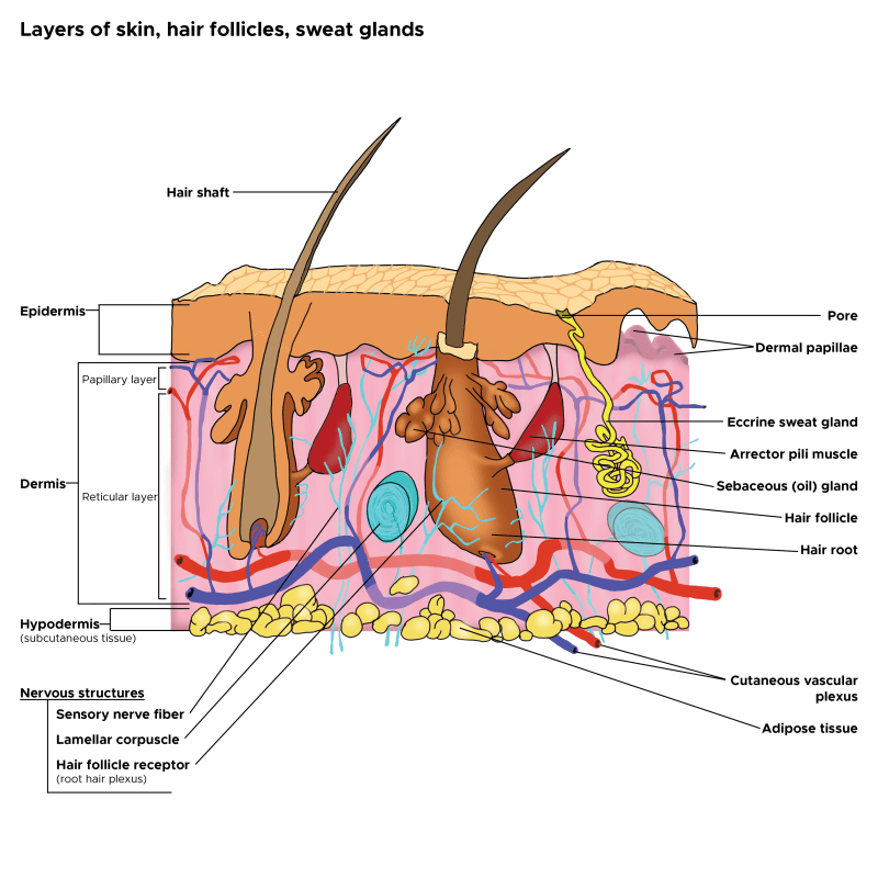
Many endogenous and exogenous factors are known to cause enlarged pilosebaceous pores. Such factors include sex, ageing, diet, chronic ultraviolet light exposure, comedogenic xenobiotics, acne, genetic and epigenetic predisposition, and seborrhoea. Most of these causative factors of enlarged pores, being exogenous and controlled by enironmental factors, means you can do something about it. There are procedures and topical products you can use to reduce pore size.

From: Yousef et al (2024)

Although the pathogenesis of enlarged facial pores is still not fully understood, three factors are thought to be key to the pathology: 1) high sebum production, 2) decreased skin elasticity around pores, and 3) increased hair follicle volume. Other factors, including chronic recurrent acne, diet, sex hormones, and skin care regimens, such as inappropriate use of cosmetics, modern Western diets, washing habits, and sun exposure, also affect pore enlargement. Many of these factors will affect the epigenetics of the skin and therefore the skin’s health and potentially pore size. Epigenetics are regulated by your environment, so there is much you can do to reduce enlarged pores.

Causes of Large Pore Size

In cross-sectioned images of conspicuous, enlarged pores, a strongly undulated epidermal–dermal junction was commonly observed around a pore’s opening. Areas with this feature correlated well to the areas with larger hollows and an uneven skin tone. (Sugata et al, 2007).

Recent clinical studies have confirmed the cause of facial pore size to be multifactorial. A positive correlation of pore size and number with sebum output level has been confirmed by several studies (Roh et al, 2006; Kim et al, 2013). Enlarged pores increase with age, up to 40 years, and then stabilize or only slightly increase (Jung et al, 2018). Another significant correlation was detected between skin elasticity and pore number in two independent studies suggesting that the structure of dermis could be involved in pore widening (Kim et al, 2013; Hameed et al, 2019). Other observations found pore counts were related to wrinkle severity; and the loss of Microfibril-associated glycoprotein-1 in the hair follicle/pore area with aging and photo-exposure, indicating a lack of matrix support in the dermis (Zheng et al, 2013; Jung et al, 2018).

Both epidermal and dermal structual impairments have been identified as a cause of large pores. Microscopic imaging of pores revealed inner structural changes affecting skin, including a lower density of collagen in the deeper dermis, a thicker stroma and coarser collagen fibers forming a tubular structure around the follicle, and an irregular basement membrane ultrastructure, all of which may result in an altered distribution of skin tensions (Sugata et al, 2008;  Sugiyama-Nakagiri et al, 2008; Mizukoshi and Takahashi, 2014). These ultrastructural alterations may result from inflammation, and recent data suggest inflammaging, mediated by complement activation (immune system proteins), as one of the possible inflammatory agents in the formation of enlarged facial pores (Qiu et al, 2024). Bacteria, such as Staphlacoccus aureus, infect hair follicles and pores, and the question remains, does the inflammation with this sort of infection enlarge the pore. Defects in epidermal morphology around pores have also been discovered, such as epidermis thickening and acanthosis (thickening of the stratum spinosum layer), likely indicating abnormal and possibly excessive keratinocyte proliferation ( Mizukoshi and Takahashi, 2014).

Procedures to Reuce Pore Size

Procedures, such as Micro-focused ultrasound with visualization (MFU-V), have been found to reduce pore size. MFU-V uses focused ultrasound energy to lift and tighten the skin by delivering heat to specific tissue layers beneath the skin’s surface, stimulating collagen production and causing skin tightening according to The Journal of Clinical and Aesthetic Dermatology. The visualization aspect of the procedure allows practitioners to see the underlying tissue during treatment, ensuring precise targeting and optimal results.

Topical S2RM to Reduce Pore Size – It’s Not Just the Exosome, It’s the Secretome

But are procedures needed to reduce pore size? No, the right choice of topical skin care products can significantly reduce pore size too. The secretome from adipose mesenchymal stem cells, something used in the NeoGenesis S2RM technology, significantly reduces pore size. That inflammation inducing the ultrastructual changes causing pores to enlarge can be reduced- reduce the inflammation with ADSC secretome found in the NeoGenesis S2RM technology. Remember, It’s Not Just the Exosome, It’s the Secretome that is optimal for reducing inflammation and regenerating tissue – including the tissue that constructs the pore. Changes of TEWL found that ADSC secretome can faciltate the recovery of the skin barrier function (Zhou et al, 2013), which can be explained by ADSC secretome normalizing the proliferation and migration of human primary keratinocytes as reported by Moon et al (2012). Both the epidermis (Ren et al, 2024) and dermis (Silveira et al, 2022) and hypodermis (An et al, 2021) are regenerated by ADSC secretome, with ADSC secretome containing collagen type IV needed to build the basment membrane, thereby regulating that “undulated epidermal–dermal junction” found to underly increased pore size.

I want to emphasie that inflammaging, inflammation that occurs as we age, is exposome induced. Those who eat well and live in an healthy envionment don’t suffer from inflammaging (Franck et al, 2025). As Franck et al write, “Inflammaging, as measured in this manner in these cohorts, thus appears to be largely a byproduct of industrialized lifestyles, with major variation across environments and populations.” In other words, if you live a healthy lifestyle, chronic inflammation, including inflammaging, is something you won’t suffer. This will reflect in your skin health, and your skin’s pore size.

Summary

Pore size in the skin depends on your envionment, your so-called exposome. Healthy skin is beautiful skin, including beautiful, healthy pores. Eat well to keep the skin healthy with sebum production levels normal and therefore reducing a risk factor for increased pore size. And the right choice of topical skin care products can help keep the skin healthy and pore size normal.

Synthetic Peptides are the Rage in Skincare – Too Bad Most Don’t Work Well

Scientists have known for decades about the benefits of natual peptides derived from our diets. However, topically applied synthetic peptides are easily broken down in the skin and have considerable difficulty penetratring the stratum corneum, both of which degrade their bioactivity. Some peptides have a high molecular weight, many are hydrophilic in character, and are highly susceptibility to enzymatic degradation, requiring the application of formulation technologies to improve the stability and the penetration of peptides through the skin. On the other hand, natural peptides produced by the human body have developed protection and functional mechanisms. If you’re using S2RM from NeoGenesis, you’re using these natually produced peptides. Learning lessons from mother nature, a few science-based companies have followed nature and are attempting to develop biomemetic peptides that are “protected and functional peptides.” Too bad most cosmetic companies don’t know about and don’t use these “protected and functional peptides.”

Written in the journal, Future Drug Discovery, “Peptides have traditionally been perceived as poor drug candidates due to unfavorable characteristics mainly regarding their pharmacokinetic behavior, including plasma stability, membrane permeability and circulation half-life” (Christina Lambers, 2022).

Peptides, along with exosomes, are “one of the skincare industry’s favorite ingredients right now,” Vogue reported in December 2024. At a Clinique launch event in early 2024, a physician declared peptides to be “the buzzword of the year.” The fashion of peptides seems to be at its peak hype (Fig.1): “Skincare is in its peptides era,” the beauty and fashion website Hypebae announced last month. As I am often asked about peptides, and because most of the literature for the lay public about peptides is incomplete and just plain wrong, I offer my short intro to peptides here. My blog is “sciencey” because it has to be in order not to present peptides in a manner that is not superficial and presents simplified dross.

.

From Maguire (2016)

What are peptides?

Basically, they’re short strings of amino acids (less than 50 amino acids). By contrast, proteins are long strings of amino acids. Peptides can be naturally produced or synthetically made in a laboratory. Naturally produced peptides are synthesized in the body as large precursor molecules (i.e., preproproteins) and are post-translationally (after the proprotein is made) processed and cleaved by proteases to generate their active peptide product. Synthetic peptides are typically made in the lab using a solid-phase peptide synthesis process where one amino acid at time is added to the string.

Problems with Synthetic Peptides

Synthetic peptides have many issues: off target activation of pathways that are detrimental to human health, poor targeting of beneficial pathways, poor penetration, and rapid degradation. Further, these synthetic peptides typically undergo lyophilization, a harsh freeze-druing process that disrupts their structure and further disrupts their safety and efficacy. Peptide aggregation and consequent loss of function is one of many problems in using synthetic peptides.

Further, the chemical synthesis process used to create peptides may introduce impurities or byproducts that can potentially lead to adverse reactions and toxicity issues. As stated in a journal from the American Chemical Society, “the current state of the art in peptide synthesis [synthetic peptides] involves primarily legacy technologies with use of large amounts of highly hazardous reagents and solvents and little focus on green chemistry and engineering” (Isidro-Llobet et al, 2019). Further, Varnava et al (2019) report that, “To date, the synthesis of peptides is concurrent with the production of enormous amounts of toxic waste.” In other words, the current industrial-scale peptide synthesis methods involve using substantial quantities of hazardous reagents and solvents, some of which may contaminate your topical product, while much of it pollutes the environment.

Synthetic peptides are cheap to make, and can easily be made in large quantities. That’s a plus for the compnies making them. But synthetic peptides can be problematic for therapeutic interactions that require post-translational modifications that are difficult to incorporate synthetically, such as glycosylation, for biological activity. Difficult to create disulfide linkages, important for the functional attributes of peptides and proteins. And peptides lack efficacy in biochemical pathways where the secondary or tertiary structure is critical or in making larger bioactive peptides and proteins. Natural peptides and proteins don’t suffer from these problems.

Microproteins ( you haven’t heard about these because they’re newly disovered) are Different from Peptides

You haven’t heard about these small proteins because they’re newly discovered and very difficult to identify and characterize. Microproteins are typically less than 100 amino acids (AAs) in length, but are different from peptides because they are not cleaved from a proprotein or protein. Until recently, microproteins have evaded detection because traditional genome annotation methods relied on stringent rules to distinguish protein coding RNAs versus non-coding RNAs (ncRNAs) to minimize the discovery of false positives including a minimum ORF length of 300 base pairs (bps). This ad hoc 100-codon threshold was initially selected based on the calculated probability that ORFs over 300 bps are significantly more likely to encode stable proteins. sORF-encoded microproteins have emerged as important new players in cellular biology and physiology, and they continue to be identified at high rates. To be clear, microproteins are polypeptides originating from short open reading frames (sORF) of less than a hundred codons. For a long time, they have been understudied because it is difficult to distinguish coding from non-coding sORFs. In recent years, the number of putatively translated sORFs has been narrowed down from hundred-thousands or millions to various thousands, owing to the advent of ribosome profiling and advances in bioinformatic and proteomic techniques. Therefore, efforts are now being made to include sORFs with robust translation evidence into databases such as GENCODE. The field of microproteins has since steadily grown, though it is still unclear how many functional coding sORFs exist in the human genome, and relatively few microproteins have been characterized to date. 

Microproteins are made in adipose mesenchymal stem cells (ADSCs) and likely released for therapeutic effect by the ADSCs (Bonilauri et al, 2021). Because of the past difficulty in identifying these microproteins, they are just now receiving attention for their therapeutic value. Understand, microproteins likely provide therapeutic value to the ADSC secretome, but our understanding of this is in its infancy.

Natural Peptides Drived from Diet

Lunasin is a 43-amino acid polypeptide originally discovered in soy. Research into the properties of lunasin began in 1996, when researchers at the University of California-Berkeley observed that the peptide arrested mitosis in cancer cells by binding to the cell’s chromatin and breaking the cell apart. The name of the peptide was chosen from the Tagalog word lunas, which means “cure”. Since its discovery, scientists have identified lunasin as the key to many of soy’s documented health benefits and it has been studied for various benefits, including cancer prevention, cholesterol management, anti-inflammation, skin health, and anti-aging. Lunasin exhibits different biological and chemopreventive properties including anti-inflammatory, anticarcinogenic, antioxidant and immune-modulating properties, anti-atherosclerosis, and osteoclastogenesis inhibition potential. Sounds great, right? But there’s a catch. Lunasin as part of a whole soy diet confers health benefits and is bioavailble, measured in human blood, but isolated lunasin has not shown such beneficial results. Isolated peptides don’t work as well as those peptides in their natural state. This is but one of many examples of natural peptides that are derived from a healthy diet providing benefit in their natural state, but when isolated, not so much happens.

Conjugated Peptides

The the human body, peptide conjugation is a crucial process involving the attachment of chemical entities to peptides to enhance their safety, efficacy, and pentration properties. These modifications, called post-translational modifications, can significantly improve characteristics like stability, targeting, and half-life of the peptide within the body, including the skin. Without peptide conjugation, the peptide is bare, subject to rapid degradation, and hydrophilic (meaning loves water and hates fat) so that it is repelled by the fatty nature of the stratum corneum. These are the problems with most synthetic peptides – they’re not conjugated. They’re not biomemetic but are cheap and can be easily hyped during the peak of the peptide-hype curve, i.e. peak of inflated expectation. However, synthetically conjugated peptides often don’t work well either.

For example, Palmitoyl Pentapeptide-4 is a conjugated peptide (pal-KTTKS). The molecular weight of Palmitoyl Pentapeptide-4 is 802.068 g/mol, larger than the “500 Dalton rule.” It consists of a pentapeptide (a chain of five amino acids, KTTKS) linked to a palmitoyl group, which is a fatty acid. This conjugation somewhat enhances its ability to penetrate the skin and makes it more effective as an anti-aging ingredient. However, Choi et al (2014) found that although pal-KTTKS was more stable than KTTKS, in dermal skin extract, 9.7% of pal-KTTKS remained after 120 min and 11.2% of pal-KTTKS remained at 60 min in the skin homogenate. In the epidermal skin extract, the concentration of pal-KTTKS throughout the 120-min incubation period was almost similar to its initial concentration. Lower amounts of proteolytic enzymes in the epidermal skin extract than in the dermal skin extract and the skin homogenate may account for pal-KTTKS lasting longer in the epidermal skin extract. 

Natural Proteins and Peptides Penetrate the Skin Better than Synthetic Peptides

Palmitoyl Pentapeptide-4 (PP-4), a conjugated peptide. Choi et al (2014) found only 11% of the peptide remains after 60 minutes in a homogenate of dermis – it’s broken down to be ineffective. Further, in skin permeation experiments, no detectable levels of KTTKS and pal-KTTKS (PP-4) were observed in the skin over a period of 48 h – the peptide dosen’t penetrate the skin.

Contrast this to the stem cell released molecules (Secretome) of ADSCs that do penentrate the skin and activate collagen production and a number of other beneficial physiological pathways. The secretome from ADSCs is loaded with proteins, peptides, microproteins, microRNA (not mRNA or DNA) and other beneficiary molecules. They work effectively, as mother nature intended, doing so collectively so that the mutlitude of molecules working togther, a systems therapeutic, exhibit synergistic, beneficial effects.

Summary

Synthetic peptides are all the rage now in skincare. Too bad most of them offer much hype and little or no benefit. There are some newer, more sophisticated conjugated peptides that I’m testing to determine whether they exhibit better efficacy than the current conjugated peptides. Stay tuned.

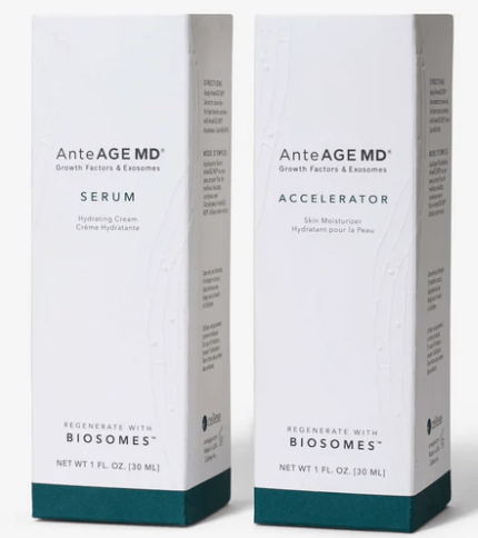
AnteAge, Founded by a Physician Whose Medical License Was Revoked and is Now Owned by Private Equity, Invents Fake Technology

John Sanderson, whose medical license was revoked for sexual misconduct and repeated negligence, has sold his company, AnteAge, to a private equity company. Now that the PE company has taken over, their marketing people have invented a new word, “Biosome.” for what is called by scientists, a “liposome.”

How do we know the Private Equity guys who own AnteAge are using fake technology? Look at the staement from their website: “Currently the AnteAGE MD bottles do not mention the new Biosome ingredient. As part of our commitment to sustainability, we have chosen to utilize our existing inner packaging rather than generating waste unnecessarily. Please rest assured that your product does in fact have Biosomes included. Please reference the ingredient listing here. Reach out with questions or refer to anteage.com.”

Here’s the statment from their web:

How Do We Know They’re Faking It

If there were actually something new in the bottle, they would have to, by law, relable the product. In other words, because they have only changed their marketing hype, and not the product’s technology, they don’t need to make any changes to the bottle labeling – specifically the bottle’s listing of ingredients. The label and the ingredients remain the same and the only thing changing is what they call the product. There’s no validation in peer-reviewed literature or patent filings confirming a unique mechanism under the name “Biosome.” Rather, it’s just marketing hype, or as some would call it, BS.

So what’s in the bottle? Liposomes. Look at the ingredients on the bottles with the new, fake technology. The list includes “Phosphatidylcholine.” Guess what are made with Phosphatidylcholine. Answer – liposomes! So now AnteAge is calling liposome, you guessed it, Biosomes. This is Private Equity at work. Say anything, do anything, for profit.

Here’s the bottle saying “Biosomes”:

And here’s the bottle’s ingredient list for the “Serum”:

Serum Ingredients:
Water (Aqua), Human Bone Marrow Stem Cell Conditioned Media, Cetyl Ethylhexanoate, Niacinamide, Dimethyl Isosorbide, Polyacrylate-13, Glycerin, Hydrolyzed Myrtus Communis Leaf Extract, Butylene Glycol,
Carbomer, Polysorbate 20, Palmitoyl Tripeptide-1, Palmitoyl, Tetrapeptide-7, Polyisobutene, Benzyl Alcohol, Salicylic Acid, Sorbic Acid, Sorbitan Isostearate, Carnosine, Ilex Paraguariensis Leaf Extract, Maltodextrin,
Disodium EDTA, DOTAP, DSPC, DSPE, DSPE PEG, Sodium Chloride, Disodium Phosphate, Potassium Phosphate, Potassium Chloride, Phosphatidylcholine, Phosphatidylserine, Sphingomyelin, Cholesterol, Mannitol,
Trehalose, sh-Oligopeptide-33, sh-Polypeptide-58, sh-Polypeptide-5, sh-Polypeptide-2, sh-Polypeptide-67, sh-Polypeptide-66, sh-Polypeptide-10, sh-Polypeptide-3, sh-Polypeptide-62, sh-Polypeptide-14,
sh-Oligopeptide-2

Bottome line. Private equity is ruining many things and now they’re lying to the public about skin care ingredients.

If you’d like to read about the science of exosomes and liposomes, you can read my 30 page academic book chapter, peer-reviewed, that I published in 2016 with Elsevier, called “Exosomes: smart nanospheres for drug delivery naturally produced by stem cells.“

Why I Don’t Formulate Products with SLS

Despite the years of research on the ill effects of SLS (sodium lauryl sulfate), I continue to hear that people, including dermatologists, are using products with this ingredient, including shampoos.

If you’ve ever Googled the causes of a skin irritation or damaged hair, you’ve likely seen posts about SLS, or sodium lauryl (or laureth) sulfate, a common ingredient in beauty products, cleansers, shampoos, toothpastes, and cleaning products.

So what does this ingredient do, why is it in everything, and what does the evidence say about how safe it is?

When we use a cleanser or shampoo, the product usually contains a detergent. That detergent is called a surfactant. A surfactant allows the oil and water molecules to bind together – it’s what’s found in soaps and detergents so we can wash our oily faces or dishes with water and remove the grime.

Sodium lauryl sulfate (SLS) is a surfactant, and its efficacy, low cost, abundance and simplicity mean it’s used in a variety of cosmetic, dermatological, and consumer products.

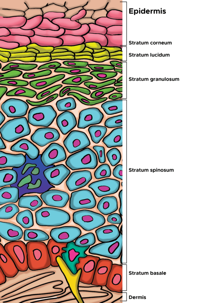
Our skin’s outermost layer, the stratum corneum of the epidermis, is specially designed to keep harmful things out, and this is where a surfactant can cause problems. Using chemicals that weaken this barrier defence mechanism can potentially cause our skin harm.

As the outermost layer of the epidermis, the stratum corneum is the first line of defense for the body, serving an essential role as a protective skin barrier against the external environment. The stratum corneum aids in hydration and water retention, which prevents skin cracking, and is made up of corneocytes, which are anucleated keratinocytes that have reached the final stage of keratinocyte differentiation (From Murphrey et al, 2022).

Some surfactants are more irritating to our skin than others. For something to be harmful, irritating or allergenic, it has to fulfill two criteria. It has to have been found in studies to irritate human skin, and it has to have the ability to penetrate the skin. SLS does both. It penetrates the stratum corneum and induces an immune reaction, and degrades the structure of the barrier.

Scientists in Germany tested 1,600 patients for SLS irritancy and found 42% of the patients tested had an irritant reaction. Another study, on seven volunteers over a three and a half month period, found regular contact caused irritation, and the irritation subsided once the skin was no longer exposed to SLS. Another study found the warmer the water used with SLS, the more irritating it will be.

SLS is a well established irritatant and is used as a positive control in dermatological testing. That is, new products being tested to see how irritating they might be to human skin are compared to the known irritant, SLS. If a person is sensitive to SLS, they might find the area that has been in contact is red, dry, scaly, itchy or sore. It’s also important to note there’s no scientific evidence SLS causes cancer, despite what is often posted on the internet. So, it’s probably OK to use SLS in products that are used for household cleaners.

Who should avoid SLS?

Everyone, especially people with a history of sensitive skin, hyperirritable skin and patients suffering from skin conditions such as atopic dermatitis (eczema), rosacea, and psoriasis are best to avoid products containing SLS. If you think it might be SLS causing a skin irritation, stop the use of the product and look for products that don’t contain SLS.

The Benefits of Topical Retinoids on the Skin

NeoGenesis uses Hydroxypinacolone retinoate (HPR), a newer retinoid that is less irritating than tretinoin and has been found to be as effective in vitro at promoting collagen production. HPR is also more stable than other retinoids in the presence of sunlight and air. Unlike retinol, HPR directly binds to the retinoid receptors and is therefore more effective and less irritating than retinol. The efficacy of HPR is similar to retinoic acid. However retinoic acid (tretinoin) can cause significant irritation of the skin, and is available only by a physician’s prescription. Retinoids can provide great benefit to aging skin. In this blog, I’ll explore some of the mechanisms by which retinoids benefit both the epidermis and the dermis. (Christine Preston contributed to this blog).

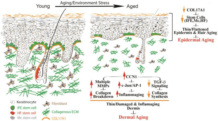
From Quan (2023) Epidermal and dermal aging of human skin. Skin aging includes the thinning of both the epidermis and dermis.

Over time, many alterations occur within the epidermis, collectively known as epidermal aging. These changes in time are characterized by the thinning of the epidermal layer and the flattening of rete ridges (as depicted in Fig 1, on the right). Rete ridges (RR) form an interdigitated surface area that reinforces cohesion between the epidermis and dermis, and this structure demonstrates plasticity, responding dynamically to stimuli such as UV irradiation. RR adapts to disruptions of its boundary during wound repair when cells lose hyper-adhesiveness, allowing the skin to appropriately remodel itself. The principal cause of epidermal aging can be traced to a reduction in the proliferation and turnover of keratinocytes, linked partially to the depletion of interfollicular epidermal (IFE) stem cells and dysfunctional Rete Ridges, leading to poor healing and thinning of the epidermis.

Collagen type, COL17A1 has been of particular interest due to its role in maintaining the homeostasis of the skin stem cells. COL17A1 is a structural element within the dermal–epidermal basement membrane, and it is synthesized by epidermal keratinocytes, not fibroblasts (Xiang et al, 2022). COL17A1 is primarily expressed in the uppermost extensions of the rete ridges area, where the niches for IFE stem cells are located. Research results have suggested a reduction in the expression of COL17A1 in human skin affected by both intrinsic and extrinsic aging factors, including human skin exposed to acute UV irradiation. The decrease in COL17A1 levels within the area specific to the rete ridges can reduce the adherence of IFE stem cells to their designated locations, leading to their removal from the skin. Consequently, the reduction of collagen protein, COL17A1, results in decreased rates of keratinocyte renewal and the development of thinner epidermal layers, the primary morphological characteristic of aging skin.

Human skin has developed two main defense mechanisms to guard against the damaging effects of UV: 1. epidermal thickening, 2. and the stimulation of melanin synthesis, however, photoprotection through increased melanogenesis is more important. As we think about retinoids and what they do for the skin, think about how retinoids help to maintain the normal structure of the skin, can actually thicken the epidermis and dermis, and how important this is for skin function and protection, including protection against UV.

Retinoids, which refer to a group of vitamin A derivatives, are among the most-extensively studied ingredients in skincare for combatting aging and enhancing the appearance of mature skin. Retinoids can stimulate collagen synthesis, inhibit MMP (Matrix metalloproteinases – too much of this activity can break-down proteinaceous tissues) activity, reduce oxidative stress, and modulate gene expression (Quan, 2023). Retinoids have exhibited efficacy in ameliorating the visual manifestations of both intrinsic and extrinsic aging, such as wrinkles, fine lines, and irregular pigmentation. The mechanisms of retinoid’s action may involve the activation of retinoic acid receptors (RARs) and retinoid X receptors (RXRs), which regulate gene transcription and cell differentiation. Retinoids may also modulate the activity of growth factors and cytokines involved in ECM turnover and inflammation. Retinoic acid (RA) is the active form of vitamin A and its precursor is called retinol (ROL). ROL can be converted into its active metabolite within human skin. When retinol is applied topically to human skin, it can penetrate the skin and undergo sequential conversion to retinaldehyde and then to retinoic acid

Skin-equivalent cultures have been used to investigate the regulatory role of retinoids in collagen homeostasis. Typically, these simplified skin constructs feature stratified and differentiated keratinocytes, representing the epidermal layer, layered atop a collagen lattice primarily comprising Type I collagen. Dermal fibroblasts are embedded within this lattice to mimic the dermal layer. When subjected to retinoic acid treatment, these skin-equivalent cultures exhibit a thickened epidermis with a substantial increase in the number of keratinocyte layers and elicit a dermal response akin to the effects observed when retinoic acid is topically applied to human skin in vivo. Consequently, skin-equivalent cultures hold significant potential as a valuable model for delving into the mechanisms by which retinoids enhance the appearance of aging skin in humans.

Increasing the Thickness of the Epidermis and the Vascularity of the Dermis in Aged Human Skin In Vivo Using Topical Retinoids: Stimulating the Growth of Epidermal Keratinocytes and Dermal Endothelial Cells

Topical application of retinoids to aged human skin in a live setting has been found to significantly enhance the thickness of the epidermis by stimulating the proliferation of epidermal keratinocytes, and increasing the number of Rete Ridges. In addition to improving epidermal thickness, topical retinoid has shown a notable increase in the proliferation of endothelial cells and blood vessels in the papillary dermis. These findings suggest that the topical application of retinoids results in the thickening of the epidermal layer and the development of fresh blood vessels within the dermis. The AP-1 transcription factor is critical to enabling the proliferation of keratinocytes in response to growth factors, cytokines, and various stimuli. The AP-1 complex consists of c-Jun and c-Fos, and it has been observed that topical retinoids significantly increases the expression of the epidermal-specific c-Jun protein, leading to a substantial increase in epidermal thickness. There is also evidence that the expression of c-Fos protein increases with retinoid treatment. These findings suggest that topical retinoids enhance the activity of the epidermal-specific c-Jun, and possibly c-Fos transcription factors, thereby stimulating the proliferation of epidermal keratinocytes in aged human skin in vivo.

Topical Retinoids Improve the Dermal ECM Microenvironment by Promoting the Production of Collagenous ECM in Aged Human Skin In Vivo

Topical retinoid treatment increases Type I collagen expression, which constitutes 80–85% of the dermal ECM, while collagen type III constitutes about 8–11%.  Topical retinoid also significantly enhances the expression of fibronectin and tropoelastin. In aged human skin in vivo, topical retinoid effectively activates dermal fibroblasts, leading to the substantial production of collagenous ECM through the activation of the TGF-β/Smad pathway, which is a key regulator of ECM production. Topical retinoid administration causes a significant increase in TGF-β1 mRNA expression and a decrease in inhibitory Smad7, while other components of the TGF-β pathway remain unaffected. Additionally, topical retinoid leads to an increase in the expression of connective tissue growth factor (CTGF/CCN2), which is substantially reduced in the dermis of aged individuals and contributes to the decline in collagen production associated with aging. These findings provide evidence that topical retinoid stimulates the production of ECM by dermal fibroblasts through the upregulation of the TGF-β/CTGF pathway in aged human skin.

In addition to the upregulation of TGF-β/CTGF pathway, retinoic acid significantly reduces CCN1 gene expression in both naturally aged and photoaged human skin in vivo. CCN1 is a negative regulator of collagen homeostasis by inhibiting the TGF-β/CTGF pathway and stimulating MMPs’ induction. These data suggest that the mechanism by which topical ROL improves aged skin, through increased collagen production and inhibition of MMPs, may involve the downregulation of CCN1. Thus, retinoids are acting through multiple pathways, inhibiting some and activating others.

In aging skin, decreased vascularity and thinning of the dermis and epidermis are substantial factors contributing to skin fragility and hindered wound healing. Blood flow to the skin, the largest organ in the body, is reduced by 40% between the ages of 20 to 70 years. Topical retinoids not only enhances ECM production, but also improves the dermal microenvironment by promoting the expansion of vasculature through endothelial cell proliferation in aged human skin. An age-related reduction in cutaneous vasculature has been reported. The increased vascularity of the dermis induced by topical retinoids can improve skin blood flow and create a more-favorable microenvironment for the homeostasis of the epidermis and dermis. Further, the promotion of epidermal keratinocyte proliferation and the restoration of ECM production by topical retinoid could create a supportive environment for the growth of endothelial cells and the development of dermal blood vessels. Epidermal keratinocytes are a significant source of vascular endothelial growth factor (VEGF), a powerful factor in promoting angiogenesis. Furthermore, increased production of dermal ECM has been demonstrated to stimulate the proliferation of endothelial cells. As a result, the augmented dermal vascularity facilitated by retinoids may have a significant impact on the homeostasis of both the epidermis and dermis.

Hydroxypinacolone Retinoate (HPR) for Anti-Aging, Photodamage, and Acne

Hydroxypinacolone retinoate (HPR) has demonstrated positive effects as a topical anti-aging ingredient, the authors of the study writing, “Together these data suggest that HPR is an effective alternative to ATRA and other less potent retinoids in the treatment of aging skin without the detrimental side-effects. And the combination of retinoids and salicylic acid can be used to ameliorate the signs of photoaging.

Data have confirmed past studies indicating that topical retinoids are under-used for acne. Further, HPR has been successfully used to treat comedonal-papular, mild to moderate acne of the face. In this study, papain was also used, in addition to HPR, as an exfoliant, and in many cases acne patients may benefit from combination therapies, such as the use of retinoids (HPR) with salicylic acid to better treat acne.

Carotenoids, Like Beta-Carotene, Convert to Retinoids When Topically Applied

You’ll notice on the label of NeoGenesis Skin Restore Serum, that in addition to HPR, carotenoids, including beta-carotene, are included in the product. While topically applied carotenoids absorb into the skin and are converted to retinoids in the skin, the carotenoids also provide antioxidant benefit to the skin. The beautiful yellow color of the vitamin A product, Skin Restore Serum, reflects the yellow pigmented carotenoid antioxidants loaded into the serum.

The amount of carotenoids in the skin depends on dietary intake, and their bioavailability from various foods, with fruits and vegetables as an important source.. After absorption in the gut and transportation into the skin, carotenoids accumulate in the skin, including the adipocytes in the hypodermis. The skin protective benefits of carotenoids, especially from those residing in the epidermis, are many, including protection from UV and air pollution.

Retinoids and Photosensitivity

Photosensitivity to retinoids appears to be a rare event, and quite to the contrary, retinoids have been found to successfully treat some forms of skin photosensitivity. First, let’s dispel the somewhat common belief that topical retinoids enhance UV-induced inflammation. Smit et al (1999) evaluated the minimal erythema dose (MED) for UVB irradiation on topical all-trans RA (tretinoin cream 0.05%) pre-treated skin compared with vehicle cream pre-treated skin and untreated skin. Their study found no significant difference for the MED values either 24 or 48 h after UVB irradiation between the all-trans RA cream treated skin, and the vehicle cream treated skin and untreated skin. In other words, topical retinoid caused no enhanced inflammation when the skin is exposed to UV.

Second, Actinic folliculitis (AF) is a rare recurrent seasonal photodermatosis, relatively newly characterized by nonpruritic, monomorphic pustules and papules appearing 4-24 h after exposure to sunlight. Lesions usually affect the face but also appear on the upper chest and arms. Resolution normally occurs within 7-10 days with cessation of sunlight exposure. AF is resistant to standard treatments used for acne vulgaris and acne rosacea, with only oral retinoids previously being reported as effective. Academic dermatologists in the UK have reported that AF responding extremely effectively to a topical retinoid.

Discussing photosensitivity, be clear that HPA is relatively stable in light and in the air. Applying and using HPA in normal lighting conditions will not degrade the product.

Long Term and Overuse of Retinoids

While a significantly higher concentration of retinol (0.4%) is required to attain similar outcomes as observed with topical retinoic acid, retinol triggers similar histological alterations (epidermal thickening and dermal ECM production) as retinoic acid. However, inappropriate or excessive use of topical retinoids or retinoic acid may also result in potential side effects. These commonly include skin dryness, redness, and peeling, which can cause discomfort. However, these side effects typically diminish over time as the skin adjusts to the product. Evidence suggests that HPR will induce fewer adverse side-effects than the other retinoids.

Long-term use of retinoids (studied for up to 2 years) have found beneficial effects to the skin throughout the treatment period, and a good safety profile. While most of the benefit is seen within 6 months following onset of the treatment, long term use can maintain the positive effects.

Summary

Topical retinoids (TR) are a safe and effective addition to one’s skin care routine, especially for aged skin. TR provides major benefits to the skin, including increased thickness of the epidermis and dermis, and enhanced blood flow to the skin. There are few side effects of retinoids, and if chosen properly, retinoid products, such as those that use HPR, are well tolerated by those with sensitive skin. Photosensitivity is not an issue, and their use with vitamin C/antioxidant products, such as those using gentle liposomal vitamin C (liposomal ascorbic acid), provides extra benefit.

Air Pollution and Eczema on the Rise

Air pollution is wreaking havoc on your skin. Simple steps to mitigate the problems associated with pollution include gentle cleansing, topical products to reduce oxidative stress and to restore barrier function.

Whether it’s jet fuel, leaded fuel for non-jet aircraft, auto exhaust, forest fires, wars such as Iraq, Gaza, and the Ukraine, or industrial fumes as causative factors, “the estimated lifetime prevalence of atopic dermatitis has increased 2~3 fold during over the past 30 years, especially in urban areas in industrialized countries, emphasizing the importance of life-style and environment in the pathogenesis of atopic diseases” (Kim, 2015). “Atopic dermatitis (AD) has increased in prevalence to become the most common inflammatory skin condition globally, and geographic variation and migration studies suggest an important role for environmental triggers” (Fadadu et al, 2023).

Air pollution can make people more sensitive to other allergens by eliciting an immune system hyper-response. This can degrade the skin’s barrier function, and now that immune system hyper-alert signaling is more exposed to the outside world and even more sensitive. Essentially, air pollution is opening up the skin by degrading it’s barrier, and making that contact between the skin’s immune system and the environment more robust. Air pollution is a catalyst in a chemical reaction, as scientists at the Max Planck Institute have written, “Fine particulate matter catalyzes oxidative stress.” The key, air pollution is causing oxidative stress in the skin, leading to chronic inflammation, a root cause in many inflammatory skin conditions, including eczema.

The illustration shows the health effects of atmospheric air pollution. The model simulations of Thomas Berkemeier and his team suggest that PM2.5 acts by conversion of a reservoir of reactive oxygen species (such as peroxides) into highly reactive OH radicals. these reactions take place in the epithelial tissue fluid, which is a thin aqueous film in the lungs and skin in which air pollutants dissolve or deposit.

For day-to-day protection from modern man’s air and water pollution and climate change (think increased fires), use air filters for the home and use mineral sunscreens with zinc or titanium, which create a physical barrier that makes it more difficult for airborne pollutants to come into direct contact with the skin. It’s also important to have a good cleansing regime. Wash the pollutants off at night with a gentle cleanser, use a product that reduces the oxidative stress, and then put on a fragrance-free hypoallergenic moisturizer containing free fatty acids, ceramide, and cholesterol that will help the skin barrier heal overnight.

Molecules Released from Human Adipose Derived Mesenchymal Stem Cells and Fibroblasts Promote Epidermal Barrier Repair

The S2RM technology that I developed is based on the molecules released from skin derived adipose mesenchymal stem cells (hASCs) and fibroblasts (HNDF). This combination of many molecules has many benefits to the skin. In this blog, I focus on the benefits of these molecules in helping to repair epidermal barrier function.

A number of diseases and conditions of the skin involve epidermal barrier dysfunction. For example, eczema is a multifactorial, heterogeneous disease associated with epidermal barrier disruption and intense systemic inflammation of the skin. Our previous work has found that S2RM attenuates the symptoms of eczema, including atopic dermatitis (AD). Studies of the mechanisms of action of the molecules present in S2RM suggest that these molecules effectively restore epidermal barrier functions in AD by facilitating the synthesis of ceramides, and creating a thicker epidermis.

hASCs as well as human dermal fibroblasts (HNDF) have a positive impact on keratinocytes proliferation, stemness maintenance, and adhesiveness to membranes via paracrine involvement when co-cultured using the collagen. This means the keratinocytes, largely responsible for building the epidermal barrier, are maintained in a younger and healthier state by the stem cells (hASCs and HNDF) that are releasing molecules into the epidermis from their location in the dermis.

These functions, along with the many other functions of the hASCs and HNDF including modulating immune function into an anti-inflammatory, pro-repair state, important to all epithelial tissues, are critical to good skin health.