Safety and Efficacy: Adipose Mesenchymal Stem Cell (ADSC) Secretome Is Superior to Bone Marrow Mesenchymal Stem Cell (BMSC) and Umbilical Cord Mesenchymal Stem Cell (UCSC) Secretomes

I list here some of the reasons why I formulate my skin care products using the secretome of adipose mesenchymal stem cells (ADSCs) instead of bone marrow mesenchymal stem cells or umbilical cord mesenchymal stem cells. ADSCs are better at reducing inflammation and setting the innate and adaptive immune systems into a pro-regenerative state, inducing collagen formation, and laying down that collagen in a manner that is anti-fibrotic. This is a small excerpt of my upcomming peer-reviewed publication.

Listing Efficacy of ADSCs Versus BMSCs versus UCMSCs Secretome (Exosomes + Soluble Fraction)

Bone Marrow Mesenchymal Stem Cells (BMSCs), and the molecules they release, prolong and enhance inflammation by increasing survival and function of neutrophils (Casatella et al, 2011; Liang et al, 2024). BMSC secretome also reprograms hematopoietic stem cells to become inflammatory white blood cells (Ng et al, 2023). Under hypoxic conditions, which induces the activation of TRL4, BMSCs secrete pro-inflammatory factors and decrease the polarization of macrophages from the M1 to M2 phenotype, the M2 type being anti-inflammatory and therefore the BMSCs are promoting more inflammation (Faulknor et al, 2017; Waterman et al, 2010). Thus, BMSCs cultured in normal hypoxic conditions in the laboratory are secreting pro-inflammatory factors and when administered to wounded skin will induce inflammation by recruiting neutrophils and M1 type pro-inflammatory macrophages.
ADSCs have consistently exhibited much greater anti‑inflammatory capabilities, phagocytic activity, anti‑apoptotic capability activity and cell viability over BMSCs (Li et al, 2019).
ADSCs have been found to be highly immunomodulating cells, exceeding the suppressive effect of BMSCs by secreting more anti-inflammatory IL-6 and transforming growth factor-β1 (TGF-β1) Ceccarelli et al (2020).
When compared with the BMSCs- and UCSCs-treated groups, the ADSCs-treated group exhibited markedly accelerated healing efficiency, characterized by increased wound closure rates, enhanced angiogenesis, and collagen deposition at the wound site in an animal model (Cao et al, 2024).
ADSCs have biological advantages over BMSCs in the proliferative capacity, secreted proteins (basic fibroblast growth factor, interferon-γ, and insulin-like growth factor-1), and immunomodulatory, ant-inflammatory effects (Li et al, 2015).
Differences in cytokine secretion cause ADSCs to have more potent immunomodulatory effects than BMSCs (Melief et al, 2013)
ADSCs are better at preventing fibrosis than BMSCs (Yoshida et al, 2023).
Adipose mesenchymal stem cell secretome is superior to that of BMSCs because it preferentially helps to rebuild the epidermis by stimulating basal keratinocytes (Ademi et al, 2023).
BMSCs express much CTHRC1 protein (Turlo et al, 2023), which may help to promote fibrosis (Liu et al, 2023).
ADSC exosomes contain SIRT1 (Huang et al, 2020) and activate SIRT1 in other cells (Liu et al, 2021) to reduce inflammation, improve mitochondrial function, and reduce senescence.
ADSC exosomes reduce inflammation in endothelial cells (Heo and Kim, 2022).
ADSCs are considered more powerful suppressors of immune response than mesenchymal stem cells (MSCs) derived from different tissue sources, including trabecular bone, bone marrow, dental pulp, and umbilical cord (Ribeiro et al., 2013; Nancarrow-Lei et al., 2017).
 ADSCs immunomodulatory effects exceed that of BMSCs (Melief et al., 2013).
ADSCs secrete higher amount of immune suppressive cytokines, such as IL-6 and transforming growth factor-β1 (TGF-β1) than do BMSCs (Soleymaninejadian et al., 2012; Melief et al., 2013; Montespan et al., 2014).
Bochev et al (2008) showed that ADSCs had a stronger ability to inhibit immunoglobulin (Ig) production by B cells than BMSCs.
Ivanova-Todorova E et al (2009) found that Adipose tissue-derived mesenchymal stem cells are more potent suppressors of the adaptive immune response through limiting dendritic cells differentiation compared to bone marrow-derived mesenchymal stem cells.
ADSC secretome inhibits LPS-induced proinflammatory cytokines (Li et al, 2018)
Human ADSCs are key regulators of immune tolerance, with the capacity to suppress T cell and inflammatory responses and to induce the generation/activation of antigen-specific regulatory T cells (Gonzalez-Rey et al, 2010).
ADSC secretome can suppress the activation, proliferation, and function of CD8+ T cells, which are inflammatory killer T cells (Kuca-Warnawin et al, 2020).
ADSC secretome was able to elevate expression of M2 macrophages and modified their cytokine expression to an anti-inflammatory profile (Hu et al, 2016; Zomer et al, 2020)
Exosomes secreted by human adipose mesenchymal stem cells promote scarless cutaneous repair by regulating extracellular matrix remodeling (Wang et al, 2017).
ADSC exosomes reduce inflammation and alleviate keloids by promoting mitochondrial autophagy through the PI3K/AKT/mTOR pathway (Liu et al, 2024).
ADSC exosomes reduce injury through the transfer of mitochondria components to neighboring cells (Xia et al, 2022).
ADSC secretome expedited wound healing and reduced inflammation in an animal model (Ma et al, 2021).
ADSC secretome promotes wound healing without leaving visible scars and was found safe when injected (An et al, 2021).
ADSC secretome has positive effects on granulation tissue formation and vascularization, and helps prevent fibrosis in pressure ulcers (Alexandrushkina et al, 2020).
Human ADSCs secrete functional neprilysin-bound exosomes that can degrade β-amyloid peptide (Aβ) that is found in the skin – cutaneous amyloidosis (Katsuda et al, 2013; Kucheryavykh et al, 2018).
In psoriasis and eczema the secretome from adipose mesenchymal stem cells (ADSCs), can regulate SOCS (suppressor of cytokine signaling) pathways, and modulate JAK pathways to reduce inflammation (Wang et al, 2022; Ko et al, 2023). Further, the secretome from ADSCs increases SOCS3 expression and, thus, the persistent and uninhibited expression of STAT3 by increased SOCS3 effectively ameliorates tissue injury by promoting tissue regeneration and decreasing inflammation and apoptosis (Lee et al, 2016).
ADSC and BMSC secretomes were characterized by the upregulation of proteins linked to ECM structure and organization and proteolytic processes compared to UCSCs, important to active involvement in tissue repair and microenvironment maintenance and suggesting their advantage for tissue-forming applications (Hodgson-Garms et al, 2025), but ADSCs are better at preventing fibrosis and reducing inflammation (Yoshida et al, 2023).
Fu et al (2025) found that hADSC-Exos are more effective in promoting hair follicle development compared to hUCMSC-Exos, and the secretome of ADSCs was more associated with growth processes such as nucleosome function than was the UCMSC secretome (Fu et al, 2025).

Mechanisms Of Action of NeoGenesis Hair Thickening Serum

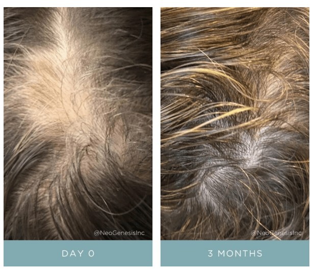
Topical application of Hair Thickening Serum (HTS) promotes hair growth by two key means: Providing, 1. Skin and hair follicle endogenous molecules from skin and hair follicle stem cells (Adipose mesenchymal stem cells, fibroblasts, and dermal papillae) that drive and maintain the transition from telogen to anagen, and 2. Botanical ingredients normally derived from healthy diets that support hair growth.

Simple topical application of NeoGenesis Hair Thickening Serum, b.i.d., twice daily.

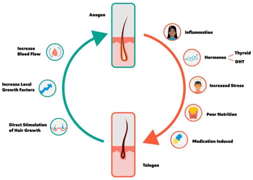
Let’s look at the hair growth cycle, and some of the many factors affecting hair growth. I’ll then explain some the mechanisms by which HTS drives the hair follicle to the anagen phase.

Figure 1. Schematic of the hair growth cycle and the factors that may influence a transition from anagen to telogen vs. telogen to anagen phase. From Natarelli et al, 2023.

HTS Mechanisms of Action in the Hair Growth Cycle

HTS’ mechanisms of action at the hair follicle are many. Here I consider a simplified summary of some of the pathways that the stem cell released molecules and botanical ingredients activate or inhibit to drive and maintain the follicle’s transition to the anagen phase.

Transition from Anagen to Telogen

Inflammation – An immunoprivileged state in the follicle is needed to drive anagen, and inflammation transitions the follicle to telogen instead (Bertolini et al, 2020). HTS reduces inflammation in the innate and adaptive immune systems by using the secretome from adipose mesenchymal stem cells – both the exosomal fraction and soluble fractions that act synergistically to optimally reduce inflammation (González-Cubero et al, 2022; Mitchell et al, 2019)

Hormone – ADSC secretome inhibits negative effects of DHT on hair growth (Tang et al, 2023; Fu et al, 2025).

Poor Nutrition – HTS contains nutrients to support hair growth. Larix Europaea Wood Extract, containing Dihydroquercetin-glucoside (polyphenol), EGCG (polyphenol catechin), glycine, zinc, Camellia Sinensis Leaf Extract, Santalum Acuminatum Fruit Extract, Citrus Glauca Fruit Extract, Acacia Victoriae Fruit Extract, Trifolium Pratense (Clover) Flower Extract (providing an abundance of polyphenols and antioxidants).

Stress – ADSC secretome mitigates immunological disturbances affecting the hair follicle (HF) and contributing to hair loss. ADSCs are able to suppress lymphocyte proliferation and, inhibit complement activation and dendritic cell differentiation from monocytes and therefore are considered natural immunosuppressants (Salhab et al, 2022).

Transition from Telogen to Anagen

Blood Flow – Secretome of ADSCs promotes angiogenesis and increased blood flow to follicles (Silveira et al, 2022; Zhu et al, 2020)

Direct stimulation of Hair Growth – Exosomes from dermal papillae cells drive hair follicle stem cell proliferation to rebuild hair follicle (Li et al, 2023), while fibroblasts provide many building-block proteins need to reconstruct the follicle architecture as it transitions from telogen to anagen (Suh et al, 2023).

Increased Local Growth factors – Fibroblasts (Lin et al, 2015), ADSCs (Won et al, 2017), and dermal papillae (HU et al, 2020) secretome all provide necessary growth factors to induce transition to anagen

References

Bertolini M et al (2020) Hair follicle immune privilege and its collapse in alopecia areata. Exp Dermatol. 29: 703–725.

Fu Y, Han YT, Xie JL, Liu RQ, Zhao B, Zhang XL, Zhang J, Zhang J. Mesenchymal stem cell exosomes enhance the development of hair follicle to ameliorate androgenetic alopecia. World J Stem Cells 2025; 17(3): 102088

Fu Y, Han YT, Xie JL, Liu RQ, Zhao B, Zhang XL, Zhang J, Zhang J. Mesenchymal stem cell exosomes enhance the development of hair follicle to ameliorate androgenetic alopecia. World J Stem Cells 2025; 17(3): 102088 [PMID: 40160691 DOI: 10.4252/wjsc.v17.i3.102088]

González-Cubero, E et al (2022) María L. González-Fernández, Elias R. Olivera, Vega Villar-Suárez,Extracellular vesicle and soluble fractions of adipose tissue-derived mesenchymal stem cells secretome induce inflammatory cytokines modulation in an in vitro model of discogenic pain,The Spine Journal,Volume 22, Issue 7,2022, Pages 1222-1234

Li J, Zhao B, Yao S, Dai Y, Zhang X, Yang N, Bao Z, Cai J, Chen Y, Wu X. Dermal PapillaCell-Derived Exosomes Regulate Hair Follicle Stem Cell Proliferation via LEF1. Int J Mol Sci. 2023 Feb 16;24(4):3961.

Lin WH, Xiang LJ, Shi HX, Zhang J, Jiang LP, Cai PT, Lin ZL, Lin BB, Huang Y, Zhang HL, Fu XB, Guo DJ, Li XK, Wang XJ, Xiao J. Fibroblast growth factors stimulate hair growth through β-catenin and Shh expression in C57BL/6 mice. Biomed Res Int. 2015;2015:730139.

Mitchell R et al (2019) Secretome of adipose-derived mesenchymal stem cells promotes skeletal muscle regeneration through synergistic action of extracellular vesicle cargo and soluble proteins. Stem Cell Res Ther. 10(1):116.

Natarelli N, Gahoonia N, Sivamani RK (2023) Integrative and Mechanistic Approach to the Hair Growth Cycle and Hair Loss. J Clin Med. 2023 Jan 23;12(3):893.

Salhab O, Khayat L, Alaaeddine N (2022) Stem cell secretome as a mechanism for restoring hair loss due to stress, particularly alopecia areata: narrative review. J Biomed Sci. 2022 Oct 5;29(1):77.

Shiqi Hu et al. (2020) Dermal exosomes containing miR-218-5p promote hair regeneration by regulating β-catenin signaling.Sci. Adv.6,eaba1685(2020).

Silveira BM, Ribeiro TO, Freitas RS, Carreira ACO, Gonçalves MS, Sogayar M, et al. (2022) Secretome from human adipose-derived mesenchymal stem cells promotes blood vessel formation and pericyte coverage in experimental skin repair. PLoS ONE 17(12): e0277863.

Suh SB, Ahn KJ, Kim EJ, Suh JY, Cho SB. (2023) Proteomic Identification and Quantification of Secretory Proteins in Human Dermal Fibroblast-Conditioned Medium for Wound Repair and Hair Regeneration. Clin Cosmet Investig Dermatol. 2023;16:1145-1157

Tang, Xin, Cao, Cuixiang, Liang, Yunxiao, Han, Le, Tu, Bin, Yu, Miao, Wan, Miaojian, Adipose-Derived Stem Cell Exosomes Antagonize the Inhibitory Effect of Dihydrotestosterone on Hair Follicle Growth by Activating Wnt/β-Catenin Pathway, Stem Cells International, 2023, 5548112, 20 pages, 2023.

Won CH et al (2017) The Basic Mechanism of Hair Growth Stimulation by Adipose-derived Stem Cells and Their Secretory Factors. Curr Stem Cell Res Ther. 2017;12(7):535-543

Zhu, D., Johnson, T.K., Wang, Y. et al. (2020) Macrophage M2 polarization induced by exosomes from adipose-derived stem cells contributes to the exosomal proangiogenic effect on mouse ischemic hindlimb. Stem Cell Res Ther 11, 162.

Why I Formulate With Chondrus Crispus Extract, and Why It’s Not Comedogenic

Chondrus crispus extract is a polysachharide, which are not comedogenic, and is known for its anti-inflammatory, moisturizing, and wound-healing properties on human skin.

NeoGenesis is a biotech company that has the most advanced skin care products on the market, utilizing, for example, our S2RM – stem cell released molecules technology (exosomes, ectosomes, and soluble fraction) that is the most advanced penetration technology in the skin care marketplace. At NeoGenesis we feature science-to-market ingredients that work and are backed by scientific and clinical studies. Chondrus crispus extract is one the science-to-market ingredients used by NeoGenesis. I’ll dig into the science of Chondrus crispus extract (CCE) in the next paragraph, but even a cursory online search of the ingredient gives you an outline of how good this extract is for the skin. Whether it’s SpecialChem, EWG, or Paula’s Choice, scientists reviewing the studies of Chondrus crispus extract all extole its virtues in skin care. Little wonder the ingredient is widely used in skin care products.

The chondrus crispus extract we use at NeoGenesis is a refined polysaccharide that has many benefits and is not comodogenic. Recent studies have found CCE mitigated inflammation and improved scratch-wound healing, and reduce environmental stress. A number of beneficial metabolites can be obtained from algae, including antioxidants, mycosporine-like amino acids, carotenoids, pigments, flavanoids, and polysaccharides. The type of polyssacharide found in chondrus crispus extract has been found to suppress oxidative stress, reduce melanogenesis, and inhibit photodamage. Further, CCE is sustainably sourced unlike a number of competing ingredients.

Iodine is an esential ingredient for skin health. Most multivitamins contain 150 mcg of iodine per daily dose, about 150 ppb. In our Barrier Renewal Cream, we use 10 parts per billion (ppb) of iodine, whcih is an extremely low concentration, representing 10 units of iodine within one billion total units. The use of iodine is being used as an active dermal agent in the treatment of inflammatory, immune-mediated and infectious diseases, e.g. psoriasis, eczema, lupus vulgaris, syphilis. Protection against UVB-induced damage and relief of inflamed skin condition has also been demonstrated (Greenwald et al, 2017).

Further, the thyroid hormone Triiodothyronine (T3) is made in the skin and requires dietary iodine to make this hormone.Control of epidermal cell production and barrier function is depenedent on this pathway (Antonini et al, 2013). Lack of iodine and thyroid hormone cause many skin conditions and loss of hair.

CCE also contains 15 of the 18 essential elements that make up the human body. This includes calcium, sulfur, magnesium, potassium, vitamin A, and vitamin K. Further, because CCE contains sulfur, it may help to reduce sebum production. CCE also contains omega-3 fatty acids, good for the skin, including acneic skin, whether topical or oral.

You can also read what other scientists and physicians say about the benefits CCE when topically applied to the skin in the popular press here, and here.

Why I Don’t Formulate Products with SLS

Despite the years of research on the ill effects of SLS (sodium lauryl sulfate), I continue to hear that people, including dermatologists, are using products with this ingredient, including shampoos.

If you’ve ever Googled the causes of a skin irritation or damaged hair, you’ve likely seen posts about SLS, or sodium lauryl (or laureth) sulfate, a common ingredient in beauty products, cleansers, shampoos, toothpastes, and cleaning products.

So what does this ingredient do, why is it in everything, and what does the evidence say about how safe it is?

When we use a cleanser or shampoo, the product usually contains a detergent. That detergent is called a surfactant. A surfactant allows the oil and water molecules to bind together – it’s what’s found in soaps and detergents so we can wash our oily faces or dishes with water and remove the grime.

Sodium lauryl sulfate (SLS) is a surfactant, and its efficacy, low cost, abundance and simplicity mean it’s used in a variety of cosmetic, dermatological, and consumer products.

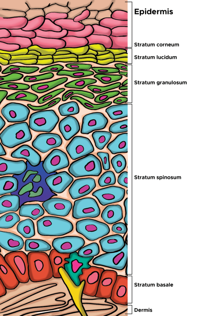
Our skin’s outermost layer, the stratum corneum of the epidermis, is specially designed to keep harmful things out, and this is where a surfactant can cause problems. Using chemicals that weaken this barrier defence mechanism can potentially cause our skin harm.

As the outermost layer of the epidermis, the stratum corneum is the first line of defense for the body, serving an essential role as a protective skin barrier against the external environment. The stratum corneum aids in hydration and water retention, which prevents skin cracking, and is made up of corneocytes, which are anucleated keratinocytes that have reached the final stage of keratinocyte differentiation (From Murphrey et al, 2022).

Some surfactants are more irritating to our skin than others. For something to be harmful, irritating or allergenic, it has to fulfill two criteria. It has to have been found in studies to irritate human skin, and it has to have the ability to penetrate the skin. SLS does both. It penetrates the stratum corneum and induces an immune reaction, and degrades the structure of the barrier.

Scientists in Germany tested 1,600 patients for SLS irritancy and found 42% of the patients tested had an irritant reaction. Another study, on seven volunteers over a three and a half month period, found regular contact caused irritation, and the irritation subsided once the skin was no longer exposed to SLS. Another study found the warmer the water used with SLS, the more irritating it will be.

SLS is a well established irritatant and is used as a positive control in dermatological testing. That is, new products being tested to see how irritating they might be to human skin are compared to the known irritant, SLS. If a person is sensitive to SLS, they might find the area that has been in contact is red, dry, scaly, itchy or sore. It’s also important to note there’s no scientific evidence SLS causes cancer, despite what is often posted on the internet. So, it’s probably OK to use SLS in products that are used for household cleaners.

Who should avoid SLS?

Everyone, especially people with a history of sensitive skin, hyperirritable skin and patients suffering from skin conditions such as atopic dermatitis (eczema), rosacea, and psoriasis are best to avoid products containing SLS. If you think it might be SLS causing a skin irritation, stop the use of the product and look for products that don’t contain SLS.

Epithelial Barrier Dysfunction in Noncommunicable and Communicable Diseases

The modern world’s dramatic increase in the number and types of chemicals in which man is exposed, a major part of of someone’s exposome, responsible for about 90% of diseases (not genetics), is causing a dramatic rise in noncommunicable and communicable diseases. Over 350 000 chemicals and mixtures of chemicals have been registered for production and use, up to three times as many as previously estimated, and an underestimate of the true number of chemical types that have been produced and commercialized. As the skin and other epithelial tissues are compromised and exposed to communicable diseases, skin and epithelial transmitted diseases are on the rise. For example, the shingles virus can enter through the skin or the epithelial tissue in our respiratory tract, and having shingles can even lead to increased risk of dementia (2nd Ref). Further, a compromised skin epithelial barrier caused by environmental factors such as mechanical trauma, exposure to exogenous proteases in microorganisms and our food, detergents, and air pollution can activate the innate and adaptive immune systems, inducing keratinocytes to release pro-inflammatory cytokines and chemokines and enhancing the antigen presentation by intradermal Langerhans cells (LCs) and dermal DCs and activating T-cells. In turn, for example, activation of T2 type T-cells leads to IL-4, IL-5, and IL-13 secretion, provoking skin barrier alteration, immune cell infiltration into skin, and itch as observed in atopic dermatitis. 

The first essential step to skin immunity is the epithelial barrier, as infection and resulting inflammation are impossible without first breaching it. Epithelia, coated with a sugary glycocalyx, not only comprise our skin but also the mucosal membranes that line our organs. Their ability to secrete squalene, mucus, lipids, and antimicrobials help protect against pathogen invasion. Additionally, epithelia can prevent inflammation by physically shoving out cells infested with toxins, allergens, antigens, pathogens, or other damage by seamlessly extruding them. This is a strategy employed by not only epithelia, but also our hair does the same as it sheds. Given that chronic inflammation could stem from a defective epithelial barrier, the current approach of treating only the inflammation will only partially mitigate symptoms of a more central problem, ongoing wound healing and disrupted barrier.

Scientists now understand that in patients with allergic disease, regardless of tissue location, the homeostatic balance of the epithelial tissue barrier is skewed toward loss of differentiation, reduced junctional integrity, and impaired innate defense and a hyperactive adaptive (trained immunity) immune system. Importantly, epithelial dysfunction characterized by these traits appears to pre-date a predisposition to immunological responses against a range of antigens or allergens, and development of allergic disease.

From the disease perspective, trained immunity is beneficial, as it improves the host’s defense against subsequent infection from pathogens. However, it can also be detrimental and result in overly active immune responses or chronic inflammation.  Even the innate immune system has some memory, given evidence that components in House Dust Mite extract activate and likely train macrophages to produce high amounts of CCL17, IL-6, and cysteinyl leukotrienes following re-exposure to HDM through the TNF-α and PGE2 pathways. Thus, an activated immune system, one that has memory and is primed to react, can lead to sensitivities that may be triggered by an overabundance of chemicals in the environment, and those sensitivities heightened by a disrupted barrier.

Evidence that epithelial barrier dysfunction explains the growing prevalence and exacerbations of inflammatory diseases such as eczema has grown through many studies performed world-wide. Diseases encompassed by the epithelial barrier theory share common features such as an increased prevalence after the 1960s that cannot be accounted soley by the emergence of improved diagnostic methods. They are indeed increasing in prevalence, i.e. the number of afflictions per 1,000 people.

Eepithelial barrier dysfunction enables the microbiome’s translocation from the skin’s surface to interepithelial and deeper subepithelial areas, doing in combination with allergens, toxins, pathogens, and pollutants. Thereafter, microbial dysbiosis and possible infection, characterized by colonization of opportunistic pathogenic bacteria and loss of the number and biodiversity of commensal bacteria results. Local inflammation, impaired tissue regeneration, and remodeling characterize the skin that suffers from impaired barrier. For example, commensal bacteria on the skin’s surface are important for epidermal lipid synthesis and improve barrier function. The skin’s microbiome is therefore critical to maintaining epidermal barrier function. The infiltration of inflammatory cells and inflammatory cytokines to affected tissues is part of the immune system’s response to erradicate invading bacteria, allergens, toxins, and pollutants away from the deep tissues. As Peter Elias, M.D. has written, “AD [atopic dermatitis] can be considered a disease of primary barrier failure, characterized by both a defective permeability (Proksch et al., 2006, and references therein) and antimicrobial function.” Further, inflammatory cells and inflammatory cytokines that migrate from the skin to other organs may play roles in the exacerbation of various inflammatory diseases in other organs. Thus, inflammation iniated in the skin may contribute to chronic inflammatory diseases in other tissues.

What Dr. Elias has been saying is that the permeability-barrier abnormality in AD is not merely an epiphenomenon but rather the “driver” of disease activity, an “outside–inside view of disease pathogenesis” (Elias and Feingold, 2001). The evidence for this is: (1) the extent of the permeability-barrier abnormality parallels severity of disease phenotype in AD, (2) both clinically uninvolved skin sites and skin cleared of inflammation for as long as 5 years continue to display significant barrier abnormalities, (3) topical artificial barrier therapy comprises effective ancillary therapy, and (4) specific replacement therapy, which targets the prominent lipid abnormalities that account for the barrier abnormality in AD, not only corrects the permeability-barrier abnormality but also comprises effective anti-inflammatory therapy for AD (Figure 1; Chamlin et al., 2002). Thus, inflammation in AD may begin with insults from without, i.e. the exposome.

That barrier insult can then activate epithelial cells in the skin, keratinocyes, which are non-professional immune cells, but do possess MHC-II molecules, that present antigens to professional immune cells, such as T-cells. Thus, with disrupted barriier, the keratinocytes can recognize antigens and present them to the immune system, leading to inflammation. More and more, scientists are discovering how epithelial cells are part of the immune system, regardless in which organ they exist. Key here is to protect barrier function in all of our epithelial tissues, including the skin.

So if inflammatory diseases such as eczema and psoriasis are environmentally triggered and lead to barrier dysfunction and resultant inflammation, what can we do?

First, calm the inflammation. It’s destructive and further degrades the epidermal barrier. S2RM technology (in NeoGenesis Recovery) is great for reducing inflammation, doing so in both the innate and adaptive immune systems.

Second, use a topical product that provides the 3 lipids and natural moisturizing factors that are needed to rebuild normal stratum corneum and barrier function. One product to use is NeoGenesis Barrier Renewal Cream (BRC).

Third, use a product that provides instantaneous barrier function and commensal bacteria. The instantaneous barrier allows the BRC to rebuld the natural barrier function over time, and the commensal bacteria help to rebuild the barrier through activation of lipid synthesis by skin cells. The commensal bacteria in Neogenesis MB-2 also help to reduce the Staphylococcus aureus infection often assicated with disrupted barrier function.

So remember, these inflammatory skin conditions are triggered by the environment. Therefore, their treatment and prevention means that if you change your environment, you can prevent or treat these diseases. Part of changing your environment is the careful choice of topical products to reduce inflammation and renormalize the structure and function of your skin.

It’s the Skin’s Architecture, Not So Much its DNA, That Causes Skin Cancer

DNA mutations in normal skin occur at high rates without cancerous growth. But when the skin’s architecture is broken down, those mutations can lead to cancer. Maintaing the skin’s architecture is critical to skin health.

Mutations Are Everywhere, But Cancer Isn’t

Scientists have looked at UV-exposed eyelid skin of middle-aged adults, and found that a square inch of normal, non-cancerous skin was riddled with mutations, many of them considered cancer drivers. The number of mutations in normal skin tissue rivaled the number seen in skin tumors, and exceeded the number of mutations seen in other tumor types, like breast cancer. Such findings once again set researchers’ expectations about how powerfully these mutations could promote cancer. There’s more to cancer than just mutations in our cells.

It’s The Architecture Stupid, Not the DNA

Prof. Dr. Cyrus Ghajar, Ph.D., a scientist at Fred Hutchinson Cancer Center, has noted that cancer-driving mutations are defined using animal studies. After identifying what is thought to be a common cancer-associated mutation in human cancers, researchers introduce the mutations into mice to see if tumors arise. If they do, they’re considered cancer drivers. But when you find these mutations in people in normal tissue, then what does that mean? It’s clearly not a driver.  Mutations, it turns out, needs partners to drive cancer. They need another powerful mutation and an abnormal microenviornment, to induce cells toward cancerous growth.

The mutation-riddled reality of normal skin tissue prompts us to realize that skin has ways of handling mutations and keeping cellular growth normal. As Prof. Dr. Mina Bissell, Ph.D. at Berkeley has taught us, our organs are set up for function, and that function is inextricably linked to chemical envionment of the cells and the architecture into which the cells are embedded. Most cells in an organ are differentiated, meaning they perform a specialized function. And this differentiated state isn’t merely governed by an internal molecular decision-making process within each cell. It’s a collective process, a top-down process, where the architecture dictates function. If a cancer cell wanders into another organ and survives, it falls under the spell of the architecture, the top-down process instructing the cancer cell to renormalize. Dr. Bissell taught us this many years ago. As shes says, “to understand cancer it is important to understand that the phenotype can override the genotype.” Further, “influences such as what you eat, your internal metabolism, inflammation and the sun’s rays” affect your phenotype and hence your genotype. For example, in the aforementioned study of eyelids, the sun is causing mutations, but the phenotype, the cellular chemistry and architecture, has overridden the genotype, the mutated DNA, and the cells are behaving normally without cancerous growth.

Cancer Reverts if Normal Architecture is Restored

Dr. Bissell and team, in a landmark study, found that if they took breast cancer cells and put them back into a normal microenvionment, a normal architecture, then the cancer cells reverted back to normal. Their results demonstrated that the extracellular matrix, i.e the architecture and its inherent chemistry, dictate the phenotype of mammary epithelial cells, and thus in the model system tested, the tissue phenotype was dominant over the cellular genotype.

A Glimpse at the Big Picture of DNA, Cells, Architecture and Downward Causation

In the big picture, what I’m talking about is downward causation. The architecture instructs the pieces what to do. So the cellular structure is instructing what the DNA, all of the DNA, needs to do. That’s downward causation. We inherit downward causation because life derives from the cell. Cells make cells. Put DNA in a dish, it sits there, inert. Put DNA into a cell, it will begin to function, with that function dependent on what cell it is in. The cell, of course, has architecture, and it is the cell’s architecture that sets boundary conditions, instructing the molecules in the cell, including the molecules in the DNA, what they should do. We humans arise from cells, the mother’s egg – and that egg receives architectural signaling from the fathers sperm, which delivers DNA contained in it own architecture, the centriole. In other words, that cellular architecture and that of it’s surroundings, is critical to the cell’s function, to creating life, and whether cells will become cancerous. Along with Dr. Mina Bissell, Prof. Dr. Dennis Nobel, Ph.D., at Oxford, has been a pioneer in this way of thinking.

Sun Exposure Can Damage the Architecture, Not Just DNA

Concerning sun exposure and skin cancer, what happens when UV damages the skin? Is DNA damaged? Yes. But damaged too is the architecture, incuding the constiuent proteins and lipids in the architecture. As Drs. Bissell and Ghajar have taught us, it’s the cells surrounding architecture that will determine whether a cell becomes cancerous. So the UV damage of the proteins and lipids that make the architecture of the skin will be critical to determing whether the skin is cancerous or normal.

What to Do to Protect the Skin’s Architecture

What do you need to do for your skin to be healthy and free from cancer? Normalize the architecture of the skin. How do you do this? 1. First, dose your skin with sunlight in moderation to protect the skin’s architecture. If you’re out for long, wear a sunblock. 2. Eat well. Fruits and vegetables contain many of the nutrients to needed to maintain and regenerate the skin’s architecture. 3. You can also utilyze a skin care routine that maintains and regenerates the skin’s architecture. Using a combination of NeoGenesis Recovery and Barrier Renewal Cream, for example, will help to maintain and regenerate the architecture of the dermis and epidermis. NeoGenesis Recovery will also help to optimize the skin’s natural ability to repair DNA. Also available are retinoid products and antioxidant skin care products that can also help to prevent damage and rebuild the skin’s architecture.

DNA Damage Repair: Animals Do It Better Than Plants and Microorganisms

Although plants and microorganisms such as algae possess some of the DNA damage response factors that are present in animal systems, they are missing many of the important regulators, such as the p53 tumor suppressor. The p53 mechanism halts the cell cycle when DNA damage is detected, giving repair machineries time to act. These observations point to the differences in the DNA damage response mechanisms between plants and animals. While DNA repair enzymes from plants may help the skin when topically applied, optimizing the DNA repair mechanisms inherent in animals is the optimal strategy for repairing DNA in animals. Adipose mesenchymal stem cell released molecules (secretome) help to optimize inherent DNA repair mechanisms when topically applied to the skin, including heat shock proteins. More importantly, the molecules help to rebuild the architecture of the skin, which prevents mutations from forming cancerous cells (see my Blog on this topic).

All plant extract contain DNA repair enzymes

If you’re using a product with plant extract, you’re likely using DNA repair enzymes. All, or nearly all, plants contain DNA repair enzymes. Keep in mind, this is important, an intrinsic feature of plant and microorganism DNA repair pathways is that they are not error-free, leading to potentially transmissible mutational alterations. The error-prone nature of some DNA repair mechanisms, however, increases the genetic diversity and variability of the populations, thus contributing to the evolution of plant genomes. In other words, despite the recent hype about DNA repair enzymes in plants/microorganisms, they don’t work well and are “error prone.”

Animals have more robust DNA reair enzymes than do plants

This is not true in animals. They are not error prone. Because animals must better protect their DNA than do plants to prevent and repair mutations that are harmeful, potentially lethal. This is why stem cells in the skin faciltate DNA repair as only animals can do.

Stem cells in the skin have the most robust DNA repair mechanisms

As stated in a presitigious scientific journal, Molecular Cell, ” adult stem cells are endowed with a superior capacity to prevent the accumulation of genetic lesions, repair them, or avoid their propagation to daughter cells, which would be particularly detrimental to the whole organism.” Further stated, “SCs [stem cells] count upon robust antioxidant defenses (which limit genotoxicity) and a superior DDR (to repair unavoidable damage). These mechanisms are in place to protect cellular homeostasis.” 

DNA repair mechanisms: it’s complicated and very efficient if enabled

DNA double strand breaks (DSBs) are a serious threat to genome stability and the erroneous repair of DNA may lead to chromosomal rearrangements with potentially lethal consequences, including cancer, for an organism. The response to DSBs elicits a highly complex and organized cellular program, called the DNA damage response (DDR), setting in motion processes that mitigate the adverse effects of DNA damage and facilitate DNA repair. Broken DNA is usually repaired by two mechanistically distinct pathways: homologous recombination (HR) and non-homologous end joining (NHEJ). HR is a complex, multistep process that allows large sections of DNA to move from one chromosome to another. NHEJ is a DNA repair mechanism that fuses broken DNA ends together without the need for a homologous template While HR uses a homologous DNA strand as a template for error-free repair, NHEJ is inherently error-prone and does not rely on sequence homology. The preferred mode of repair and cellular consequences of DDR varies between organisms and is also dependant on cell type and cell cycle context. For example, while HR is the preferred mode of repair in many unicellular organisms such as budding and fission yeast, NHEJ is the prevalent pathway in plants and animals.

Plants versus animals, digging deeper into the mechanisms

However, in many aspects, plants respond differently to DNA insults than do animals. The constant risk of tumor formation in animals has led to evolution of DDR that assures precise genome maintenance, often resulting in apoptotic death of significantly damaged cells. The lack of such a strong selective constraint presumably permitted evolution of a less potent DDR in plants, making plants more prone to genome damage. Furthermore, plant cells are exposed to high levels of genotoxic stress resulting from long-term exposure to solar ultraviolet (UV) irradiation, photosynthesis and extended periods of desiccation. Thus, some features of plant DDR and DSB repair may deviate from models primarily established from studies in yeasts and mammals.

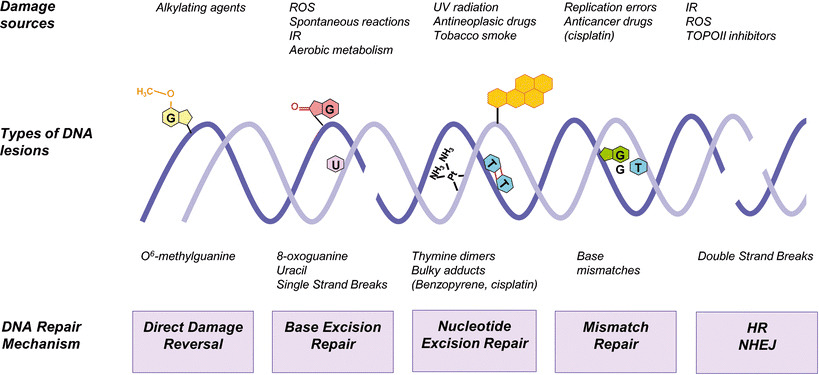
DNA repair in animals is even more complicated than that described by the two major pathways. Five DNA repair mechanisms are usually distinguished: (a) direct DNA damage reversal, (b) BER, (c) mismatch repair (MMR), (d) nucleotide excision repair (NER), and (e) homologous recombination (HR) and non-homologous end joining (NHEJ). DNA repair pathways were originally restricted to the nuclear compartment. Ample evidence indicates that mitochondria possess a number of DNA repair factors and mechanisms shared with the nuclear processes.

From: Sottile and Nadin (2018). Sources of DNA damage and repair mechanisms. Endogenous and exogenous agents constantly impact on DNA. They may cause many different forms of DNA damage. The scheme shows the five major DNA repair mechanisms operating in the nucleus of mammalian cells capable of removing a wide range of DNA lesions: direct damage reversal, base excision repair (BER), nucleotide excision repair (NER), mismatch repair (MMR), homologous recombination (HR), and non-homologous end joining (NHEJ). The BER system may also be found in the mitochondria. ROS reactive oxygen species, IR ionizing radiation, TOPOII topoisomerase II

Not included in the above summary is a new form of DNA repair in animals, called neucleophagy. This was just reported in October 2024 by a group of scientists at Oxford. Found to be evolutionarily conserved and clinically relevant, we’ll know more baout this mechanism in the coming years. Its mediated by TEX264, an intrinsically disordered protein, and as such, I suspect this will be an important and widespread DNA repair mechanism, possibly involving adult stem cells and their secretome – stay tuned.

How adult adipose mesenchymal stem cell scretome faciltates DNA repair

These complicated processes are faciltated by a number of molecules, including proteins, such as heat shock proteins, released by adipose mesenchymal stem cells (ADSCs). Another example, ADSCs release sirtuins, which are involved in DNA repair. The sirtuins work both as protein activators and chromatin-structure-modifying enzymes. Deacetylation carried by sirtuins represents a basic epigenetic mechanism. Histone modifications including deacetylation and poly-(ADP)-ribosylation compromise an essential part of physiological ageing processes that are involved in the pathogenesis of ageing-related diseases. Stem cells are also known to produce 5-hydroxymethylcytosine binding, embryonic stem-cell-specific (HMCES) protein functions as an intermediate in DNA interstrand cross-link repair, part of BER. Antioxidants from ADSCs are also important for DNA repair. They include: Superoxide dismutase (SOD): This enzyme converts superoxide radicals (O2-) into hydrogen peroxide (H2O2), which is then further broken down by other enzymes like catalase. Catalase (CAT): Catalase directly breaks down hydrogen peroxide (H2O2) into water and oxygen, preventing further oxidative damage to cellular components including DNA.  Peroxiredoxins (Prxs): This family of enzymes also plays a significant role in scavenging reactive oxygen species, particularly in the nucleus where DNA is located, and can directly contribute to DNA repair mechanisms.

The secretome from ADSCs was tested for skin repair following irradiation, where DNA and protein damage is a key component in the radiation dermatitis. Working with Dr, Michael Traub, N.D., NeoGeneis found that simple topical application of ADSC secretome in its Recovery product significantly reduced radiation dermatitis. The Recovery works through the many types of molecules found in the ADSC (and fibroblast) secretome that enable the skin’s robust DNA repair mechanisms to work optimally.

Beyond the hype of plant/microorganism DNA repair enzymes

You can see that using the secretome from ADSCs is much more powerful than using a plant or microorganism extract to repair DNA. While DNA repair enzymes from plants and microorganisms have been found to reduce cyclobutane pyrimidine dimers, and presumably repair DNA in humans when compared to doing nothing, the “DNA repair” products on the market contain many other ingredients that are at least partially responsible for their efficacy. Those other ingredients include antioxidants and sunblocks. If you want a great topical product to repair DNA in your skin cells, use NeoGenesis Recovery that is full of the molecules released from ADSCs, including those molecules in exosomes and those molecules in the soluble fraction.

The Benefits of Topical Retinoids on the Skin

NeoGenesis uses Hydroxypinacolone retinoate (HPR), a newer retinoid that is less irritating than tretinoin and has been found to be as effective in vitro at promoting collagen production. HPR is also more stable than other retinoids in the presence of sunlight and air. Unlike retinol, HPR directly binds to the retinoid receptors and is therefore more effective and less irritating than retinol. The efficacy of HPR is similar to retinoic acid. However retinoic acid (tretinoin) can cause significant irritation of the skin, and is available only by a physician’s prescription. Retinoids can provide great benefit to aging skin. In this blog, I’ll explore some of the mechanisms by which retinoids benefit both the epidermis and the dermis. (Christine Preston contributed to this blog).

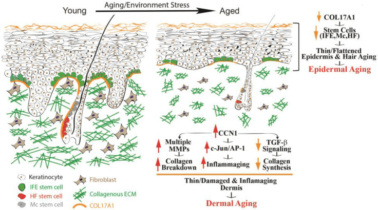
From Quan (2023) Epidermal and dermal aging of human skin. Skin aging includes the thinning of both the epidermis and dermis.

Over time, many alterations occur within the epidermis, collectively known as epidermal aging. These changes in time are characterized by the thinning of the epidermal layer and the flattening of rete ridges (as depicted in Fig 1, on the right). Rete ridges (RR) form an interdigitated surface area that reinforces cohesion between the epidermis and dermis, and this structure demonstrates plasticity, responding dynamically to stimuli such as UV irradiation. RR adapts to disruptions of its boundary during wound repair when cells lose hyper-adhesiveness, allowing the skin to appropriately remodel itself. The principal cause of epidermal aging can be traced to a reduction in the proliferation and turnover of keratinocytes, linked partially to the depletion of interfollicular epidermal (IFE) stem cells and dysfunctional Rete Ridges, leading to poor healing and thinning of the epidermis.

Collagen type, COL17A1 has been of particular interest due to its role in maintaining the homeostasis of the skin stem cells. COL17A1 is a structural element within the dermal–epidermal basement membrane, and it is synthesized by epidermal keratinocytes, not fibroblasts (Xiang et al, 2022). COL17A1 is primarily expressed in the uppermost extensions of the rete ridges area, where the niches for IFE stem cells are located. Research results have suggested a reduction in the expression of COL17A1 in human skin affected by both intrinsic and extrinsic aging factors, including human skin exposed to acute UV irradiation. The decrease in COL17A1 levels within the area specific to the rete ridges can reduce the adherence of IFE stem cells to their designated locations, leading to their removal from the skin. Consequently, the reduction of collagen protein, COL17A1, results in decreased rates of keratinocyte renewal and the development of thinner epidermal layers, the primary morphological characteristic of aging skin.

Human skin has developed two main defense mechanisms to guard against the damaging effects of UV: 1. epidermal thickening, 2. and the stimulation of melanin synthesis, however, photoprotection through increased melanogenesis is more important. As we think about retinoids and what they do for the skin, think about how retinoids help to maintain the normal structure of the skin, can actually thicken the epidermis and dermis, and how important this is for skin function and protection, including protection against UV.

Retinoids, which refer to a group of vitamin A derivatives, are among the most-extensively studied ingredients in skincare for combatting aging and enhancing the appearance of mature skin. Retinoids can stimulate collagen synthesis, inhibit MMP (Matrix metalloproteinases – too much of this activity can break-down proteinaceous tissues) activity, reduce oxidative stress, and modulate gene expression (Quan, 2023). Retinoids have exhibited efficacy in ameliorating the visual manifestations of both intrinsic and extrinsic aging, such as wrinkles, fine lines, and irregular pigmentation. The mechanisms of retinoid’s action may involve the activation of retinoic acid receptors (RARs) and retinoid X receptors (RXRs), which regulate gene transcription and cell differentiation. Retinoids may also modulate the activity of growth factors and cytokines involved in ECM turnover and inflammation. Retinoic acid (RA) is the active form of vitamin A and its precursor is called retinol (ROL). ROL can be converted into its active metabolite within human skin. When retinol is applied topically to human skin, it can penetrate the skin and undergo sequential conversion to retinaldehyde and then to retinoic acid

Skin-equivalent cultures have been used to investigate the regulatory role of retinoids in collagen homeostasis. Typically, these simplified skin constructs feature stratified and differentiated keratinocytes, representing the epidermal layer, layered atop a collagen lattice primarily comprising Type I collagen. Dermal fibroblasts are embedded within this lattice to mimic the dermal layer. When subjected to retinoic acid treatment, these skin-equivalent cultures exhibit a thickened epidermis with a substantial increase in the number of keratinocyte layers and elicit a dermal response akin to the effects observed when retinoic acid is topically applied to human skin in vivo. Consequently, skin-equivalent cultures hold significant potential as a valuable model for delving into the mechanisms by which retinoids enhance the appearance of aging skin in humans.

Increasing the Thickness of the Epidermis and the Vascularity of the Dermis in Aged Human Skin In Vivo Using Topical Retinoids: Stimulating the Growth of Epidermal Keratinocytes and Dermal Endothelial Cells

Topical application of retinoids to aged human skin in a live setting has been found to significantly enhance the thickness of the epidermis by stimulating the proliferation of epidermal keratinocytes, and increasing the number of Rete Ridges. In addition to improving epidermal thickness, topical retinoid has shown a notable increase in the proliferation of endothelial cells and blood vessels in the papillary dermis. These findings suggest that the topical application of retinoids results in the thickening of the epidermal layer and the development of fresh blood vessels within the dermis. The AP-1 transcription factor is critical to enabling the proliferation of keratinocytes in response to growth factors, cytokines, and various stimuli. The AP-1 complex consists of c-Jun and c-Fos, and it has been observed that topical retinoids significantly increases the expression of the epidermal-specific c-Jun protein, leading to a substantial increase in epidermal thickness. There is also evidence that the expression of c-Fos protein increases with retinoid treatment. These findings suggest that topical retinoids enhance the activity of the epidermal-specific c-Jun, and possibly c-Fos transcription factors, thereby stimulating the proliferation of epidermal keratinocytes in aged human skin in vivo.

Topical Retinoids Improve the Dermal ECM Microenvironment by Promoting the Production of Collagenous ECM in Aged Human Skin In Vivo

Topical retinoid treatment increases Type I collagen expression, which constitutes 80–85% of the dermal ECM, while collagen type III constitutes about 8–11%.  Topical retinoid also significantly enhances the expression of fibronectin and tropoelastin. In aged human skin in vivo, topical retinoid effectively activates dermal fibroblasts, leading to the substantial production of collagenous ECM through the activation of the TGF-β/Smad pathway, which is a key regulator of ECM production. Topical retinoid administration causes a significant increase in TGF-β1 mRNA expression and a decrease in inhibitory Smad7, while other components of the TGF-β pathway remain unaffected. Additionally, topical retinoid leads to an increase in the expression of connective tissue growth factor (CTGF/CCN2), which is substantially reduced in the dermis of aged individuals and contributes to the decline in collagen production associated with aging. These findings provide evidence that topical retinoid stimulates the production of ECM by dermal fibroblasts through the upregulation of the TGF-β/CTGF pathway in aged human skin.

In addition to the upregulation of TGF-β/CTGF pathway, retinoic acid significantly reduces CCN1 gene expression in both naturally aged and photoaged human skin in vivo. CCN1 is a negative regulator of collagen homeostasis by inhibiting the TGF-β/CTGF pathway and stimulating MMPs’ induction. These data suggest that the mechanism by which topical ROL improves aged skin, through increased collagen production and inhibition of MMPs, may involve the downregulation of CCN1. Thus, retinoids are acting through multiple pathways, inhibiting some and activating others.

In aging skin, decreased vascularity and thinning of the dermis and epidermis are substantial factors contributing to skin fragility and hindered wound healing. Blood flow to the skin, the largest organ in the body, is reduced by 40% between the ages of 20 to 70 years. Topical retinoids not only enhances ECM production, but also improves the dermal microenvironment by promoting the expansion of vasculature through endothelial cell proliferation in aged human skin. An age-related reduction in cutaneous vasculature has been reported. The increased vascularity of the dermis induced by topical retinoids can improve skin blood flow and create a more-favorable microenvironment for the homeostasis of the epidermis and dermis. Further, the promotion of epidermal keratinocyte proliferation and the restoration of ECM production by topical retinoid could create a supportive environment for the growth of endothelial cells and the development of dermal blood vessels. Epidermal keratinocytes are a significant source of vascular endothelial growth factor (VEGF), a powerful factor in promoting angiogenesis. Furthermore, increased production of dermal ECM has been demonstrated to stimulate the proliferation of endothelial cells. As a result, the augmented dermal vascularity facilitated by retinoids may have a significant impact on the homeostasis of both the epidermis and dermis.

Hydroxypinacolone Retinoate (HPR) for Anti-Aging, Photodamage, and Acne

Hydroxypinacolone retinoate (HPR) has demonstrated positive effects as a topical anti-aging ingredient, the authors of the study writing, “Together these data suggest that HPR is an effective alternative to ATRA and other less potent retinoids in the treatment of aging skin without the detrimental side-effects. And the combination of retinoids and salicylic acid can be used to ameliorate the signs of photoaging.

Data have confirmed past studies indicating that topical retinoids are under-used for acne. Further, HPR has been successfully used to treat comedonal-papular, mild to moderate acne of the face. In this study, papain was also used, in addition to HPR, as an exfoliant, and in many cases acne patients may benefit from combination therapies, such as the use of retinoids (HPR) with salicylic acid to better treat acne.

Carotenoids, Like Beta-Carotene, Convert to Retinoids When Topically Applied

You’ll notice on the label of NeoGenesis Skin Restore Serum, that in addition to HPR, carotenoids, including beta-carotene, are included in the product. While topically applied carotenoids absorb into the skin and are converted to retinoids in the skin, the carotenoids also provide antioxidant benefit to the skin. The beautiful yellow color of the vitamin A product, Skin Restore Serum, reflects the yellow pigmented carotenoid antioxidants loaded into the serum.

The amount of carotenoids in the skin depends on dietary intake, and their bioavailability from various foods, with fruits and vegetables as an important source.. After absorption in the gut and transportation into the skin, carotenoids accumulate in the skin, including the adipocytes in the hypodermis. The skin protective benefits of carotenoids, especially from those residing in the epidermis, are many, including protection from UV and air pollution.

Retinoids and Photosensitivity

Photosensitivity to retinoids appears to be a rare event, and quite to the contrary, retinoids have been found to successfully treat some forms of skin photosensitivity. First, let’s dispel the somewhat common belief that topical retinoids enhance UV-induced inflammation. Smit et al (1999) evaluated the minimal erythema dose (MED) for UVB irradiation on topical all-trans RA (tretinoin cream 0.05%) pre-treated skin compared with vehicle cream pre-treated skin and untreated skin. Their study found no significant difference for the MED values either 24 or 48 h after UVB irradiation between the all-trans RA cream treated skin, and the vehicle cream treated skin and untreated skin. In other words, topical retinoid caused no enhanced inflammation when the skin is exposed to UV.

Second, Actinic folliculitis (AF) is a rare recurrent seasonal photodermatosis, relatively newly characterized by nonpruritic, monomorphic pustules and papules appearing 4-24 h after exposure to sunlight. Lesions usually affect the face but also appear on the upper chest and arms. Resolution normally occurs within 7-10 days with cessation of sunlight exposure. AF is resistant to standard treatments used for acne vulgaris and acne rosacea, with only oral retinoids previously being reported as effective. Academic dermatologists in the UK have reported that AF responding extremely effectively to a topical retinoid.

Discussing photosensitivity, be clear that HPA is relatively stable in light and in the air. Applying and using HPA in normal lighting conditions will not degrade the product.

Long Term and Overuse of Retinoids

While a significantly higher concentration of retinol (0.4%) is required to attain similar outcomes as observed with topical retinoic acid, retinol triggers similar histological alterations (epidermal thickening and dermal ECM production) as retinoic acid. However, inappropriate or excessive use of topical retinoids or retinoic acid may also result in potential side effects. These commonly include skin dryness, redness, and peeling, which can cause discomfort. However, these side effects typically diminish over time as the skin adjusts to the product. Evidence suggests that HPR will induce fewer adverse side-effects than the other retinoids.

Long-term use of retinoids (studied for up to 2 years) have found beneficial effects to the skin throughout the treatment period, and a good safety profile. While most of the benefit is seen within 6 months following onset of the treatment, long term use can maintain the positive effects.

Summary

Topical retinoids (TR) are a safe and effective addition to one’s skin care routine, especially for aged skin. TR provides major benefits to the skin, including increased thickness of the epidermis and dermis, and enhanced blood flow to the skin. There are few side effects of retinoids, and if chosen properly, retinoid products, such as those that use HPR, are well tolerated by those with sensitive skin. Photosensitivity is not an issue, and their use with vitamin C/antioxidant products, such as those using gentle liposomal vitamin C (liposomal ascorbic acid), provides extra benefit.