Skin Pores – Reducing Their Size

Many endogenous and exogenous factors are known to cause enlarged pilosebaceous pores. Such factors include sex, ageing, diet, chronic ultraviolet light exposure, comedogenic xenobiotics, acne, genetic and epigenetic predisposition, and seborrhoea. Most of these causative factors of enlarged pores, being exogenous and controlled by enironmental factors, means you can do something about it. There are procedures and topical products you can use to reduce pore size.

From: Yousef et al (2024)

Although the pathogenesis of enlarged facial pores is still not fully understood, three factors are thought to be key to the pathology: 1) high sebum production, 2) decreased skin elasticity around pores, and 3) increased hair follicle volume. Other factors, including chronic recurrent acne, diet, sex hormones, and skin care regimens, such as inappropriate use of cosmetics, modern Western diets, washing habits, and sun exposure, also affect pore enlargement. Many of these factors will affect the epigenetics of the skin and therefore the skin’s health and potentially pore size. Epigenetics are regulated by your environment, so there is much you can do to reduce enlarged pores.

Causes of Large Pore Size

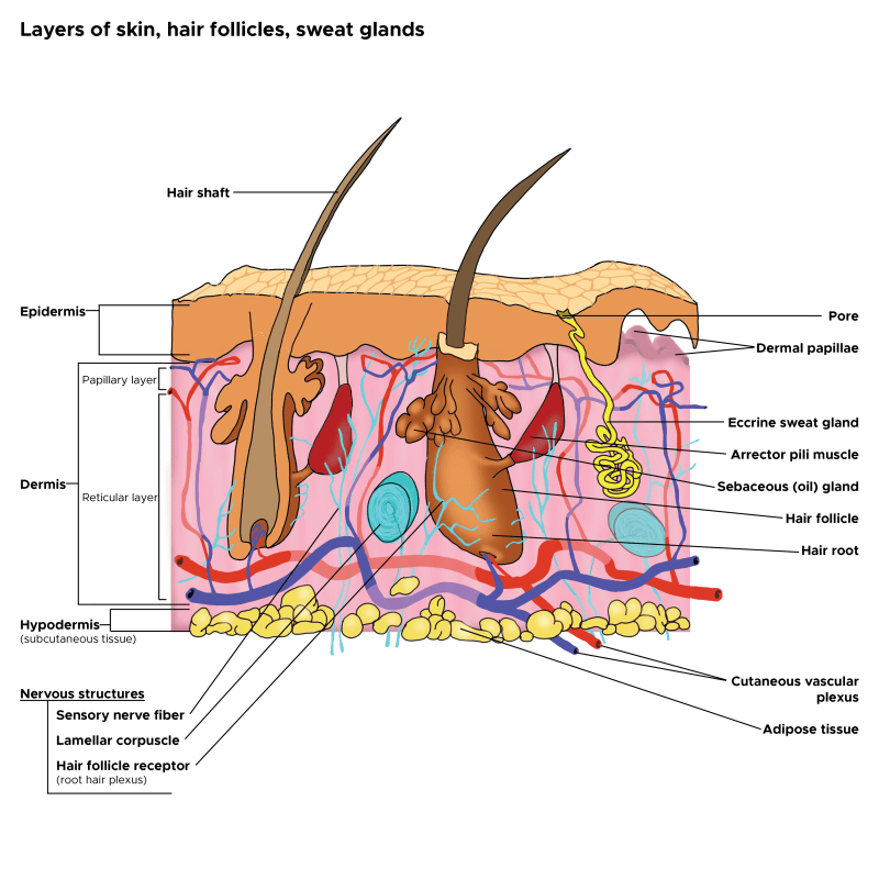
In cross-sectioned images of conspicuous, enlarged pores, a strongly undulated epidermal–dermal junction was commonly observed around a pore’s opening. Areas with this feature correlated well to the areas with larger hollows and an uneven skin tone. (Sugata et al, 2007).

Recent clinical studies have confirmed the cause of facial pore size to be multifactorial. A positive correlation of pore size and number with sebum output level has been confirmed by several studies (Roh et al, 2006Kim et al, 2013). Enlarged pores increase with age, up to 40 years, and then stabilize or only slightly increase (Jung et al, 2018). Another significant correlation was detected between skin elasticity and pore number in two independent studies suggesting that the structure of dermis could be involved in pore widening (Kim et al, 2013; Hameed et al, 2019). Other observations found pore counts were related to wrinkle severity; and the loss of Microfibril-associated glycoprotein-1 in the hair follicle/pore area with aging and photo-exposure, indicating a lack of matrix support in the dermis (Zheng et al, 2013; Jung et al, 2018).

Both epidermal and dermal structual impairments have been identified as a cause of large pores. Microscopic imaging of pores revealed inner structural changes affecting skin, including a lower density of collagen in the deeper dermis, a thicker stroma and coarser collagen fibers forming a tubular structure around the follicle, and an irregular basement membrane ultrastructure, all of which may result in an altered distribution of skin tensions (Sugata et al, 2008;  Sugiyama-Nakagiri et al, 2008; Mizukoshi and Takahashi, 2014). These ultrastructural alterations may result from inflammation, and recent data suggest inflammaging, mediated by complement activation (immune system proteins), as one of the possible inflammatory agents in the formation of enlarged facial pores (Qiu et al, 2024). Bacteria, such as Staphlacoccus aureus, infect hair follicles and pores, and the question remains, does the inflammation with this sort of infection enlarge the pore. Defects in epidermal morphology around pores have also been discovered, such as epidermis thickening and acanthosis (thickening of the stratum spinosum layer), likely indicating abnormal and possibly excessive keratinocyte proliferation ( Mizukoshi and Takahashi, 2014).

Procedures to Reuce Pore Size

Procedures, such as Micro-focused ultrasound with visualization (MFU-V), have been found to reduce pore size. MFU-V uses focused ultrasound energy to lift and tighten the skin by delivering heat to specific tissue layers beneath the skin’s surface, stimulating collagen production and causing skin tightening according to The Journal of Clinical and Aesthetic Dermatology. The visualization aspect of the procedure allows practitioners to see the underlying tissue during treatment, ensuring precise targeting and optimal results.

Topical S2RM to Reduce Pore Size It’s Not Just the Exosome, It’s the Secretome

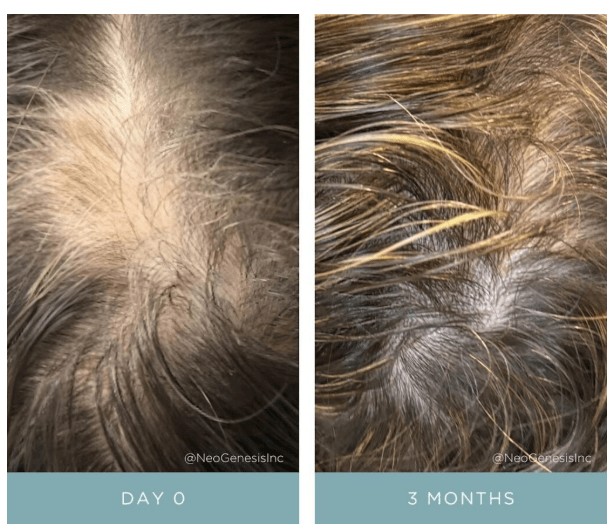
But are procedures needed to reduce pore size? No, the right choice of topical skin care products can significantly reduce pore size too. The secretome from adipose mesenchymal stem cells, something used in the NeoGenesis S2RM technology, significantly reduces pore size. That inflammation inducing the ultrastructual changes causing pores to enlarge can be reduced- reduce the inflammation with ADSC secretome found in the NeoGenesis S2RM technology. Remember, It’s Not Just the Exosome, It’s the Secretome that is optimal for reducing inflammation and regenerating tissue – including the tissue that constructs the pore. Changes of TEWL found that ADSC secretome can faciltate the recovery of the skin barrier function (Zhou et al, 2013), which can be explained by ADSC secretome normalizing the proliferation and migration of human primary keratinocytes as reported by Moon et al (2012). Both the epidermis (Ren et al, 2024) and dermis (Silveira et al, 2022) and hypodermis (An et al, 2021) are regenerated by ADSC secretome, with ADSC secretome containing collagen type IV needed to build the basment membrane, thereby regulating that “undulated epidermal–dermal junction” found to underly increased pore size.

I want to emphasie that inflammaging, inflammation that occurs as we age, is exposome induced. Those who eat well and live in an healthy envionment don’t suffer from inflammaging (Franck et al, 2025). As Franck et al write, “Inflammaging, as measured in this manner in these cohorts, thus appears to be largely a byproduct of industrialized lifestyles, with major variation across environments and populations.” In other words, if you live a healthy lifestyle, chronic inflammation, including inflammaging, is something you won’t suffer. This will reflect in your skin health, and your skin’s pore size.

Summary

Pore size in the skin depends on your envionment, your so-called exposome. Healthy skin is beautiful skin, including beautiful, healthy pores. Eat well to keep the skin healthy with sebum production levels normal and therefore reducing a risk factor for increased pore size. And the right choice of topical skin care products can help keep the skin healthy and pore size normal.

Safety and Efficacy: Adipose Mesenchymal Stem Cell (ADSC) Secretome Is Superior to Bone Marrow Mesenchymal Stem Cell (BMSC) and Umbilical Cord Mesenchymal Stem Cell (UCSC) Secretomes

I list here some of the reasons why I formulate my skin care products using the secretome of adipose mesenchymal stem cells (ADSCs) instead of bone marrow mesenchymal stem cells or umbilical cord mesenchymal stem cells. ADSCs are better at reducing inflammation and setting the innate and adaptive immune systems into a pro-regenerative state, inducing collagen formation, and laying down that collagen in a manner that is anti-fibrotic. This is a small excerpt of my upcomming peer-reviewed publication.

Listing Efficacy of ADSCs Versus BMSCs versus UCMSCs Secretome (Exosomes + Soluble Fraction)

Bone Marrow Mesenchymal Stem Cells (BMSCs), and the molecules they release, prolong and enhance inflammation by increasing survival and function of neutrophils (Casatella et al, 2011; Liang et al, 2024). BMSC secretome also reprograms hematopoietic stem cells to become inflammatory white blood cells (Ng et al, 2023). Under hypoxic conditions, which induces the activation of TRL4, BMSCs secrete pro-inflammatory factors and decrease the polarization of macrophages from the M1 to M2 phenotype, the M2 type being anti-inflammatory and therefore the BMSCs are promoting more inflammation (Faulknor et al, 2017; Waterman et al, 2010). Thus, BMSCs cultured in normal hypoxic conditions in the laboratory are secreting pro-inflammatory factors and when administered to wounded skin will induce inflammation by recruiting neutrophils and M1 type pro-inflammatory macrophages.
ADSCs have consistently exhibited much greater anti‑inflammatory capabilities, phagocytic activity, anti‑apoptotic capability activity and cell viability over BMSCs (Li et al, 2019).
ADSCs have been found to be highly immunomodulating cells, exceeding the suppressive effect of BMSCs by secreting more anti-inflammatory IL-6 and transforming growth factor-β1 (TGF-β1) Ceccarelli et al (2020).
When compared with the BMSCs- and UCSCs-treated groups, the ADSCs-treated group exhibited markedly accelerated healing efficiency, characterized by increased wound closure rates, enhanced angiogenesis, and collagen deposition at the wound site in an animal model (Cao et al, 2024).
ADSCs have biological advantages over BMSCs in the proliferative capacity, secreted proteins (basic fibroblast growth factor, interferon-γ, and insulin-like growth factor-1), and immunomodulatory, ant-inflammatory effects (Li et al, 2015).
Differences in cytokine secretion cause ADSCs to have more potent immunomodulatory effects than BMSCs (Melief et al, 2013)
ADSCs are better at preventing fibrosis than BMSCs (Yoshida et al, 2023).
Adipose mesenchymal stem cell secretome is superior to that of BMSCs because it preferentially helps to rebuild the epidermis by stimulating basal keratinocytes (Ademi et al, 2023).
BMSCs express much CTHRC1 protein (Turlo et al, 2023), which may help to promote fibrosis (Liu et al, 2023).
ADSC exosomes contain SIRT1 (Huang et al, 2020) and activate SIRT1 in other cells (Liu et al, 2021) to reduce inflammation, improve mitochondrial function, and reduce senescence.
ADSC exosomes reduce inflammation in endothelial cells (Heo and Kim, 2022).
ADSCs are considered more powerful suppressors of immune response than mesenchymal stem cells (MSCs) derived from different tissue sources, including trabecular bone, bone marrow, dental pulp, and umbilical cord (Ribeiro et al., 2013; Nancarrow-Lei et al., 2017).
 ADSCs immunomodulatory effects exceed that of BMSCs (Melief et al., 2013).
ADSCs secrete higher amount of immune suppressive cytokines, such as IL-6 and transforming growth factor-β1 (TGF-β1) than do BMSCs (Soleymaninejadian et al., 2012; Melief et al., 2013; Montespan et al., 2014).
Bochev et al (2008) showed that ADSCs had a stronger ability to inhibit immunoglobulin (Ig) production by B cells than BMSCs.
Ivanova-Todorova E et al (2009) found that Adipose tissue-derived mesenchymal stem cells are more potent suppressors of the adaptive immune response through limiting dendritic cells differentiation compared to bone marrow-derived mesenchymal stem cells.
ADSC secretome inhibits LPS-induced proinflammatory cytokines (Li et al, 2018)
Human ADSCs are key regulators of immune tolerance, with the capacity to suppress T cell and inflammatory responses and to induce the generation/activation of antigen-specific regulatory T cells (Gonzalez-Rey et al, 2010).
ADSC secretome can suppress the activation, proliferation, and function of CD8+ T cells, which are inflammatory killer T cells (Kuca-Warnawin et al, 2020).
ADSC secretome was able to elevate expression of M2 macrophages and modified their cytokine expression to an anti-inflammatory profile (Hu et al, 2016; Zomer et al, 2020)
Exosomes secreted by human adipose mesenchymal stem cells promote scarless cutaneous repair by regulating extracellular matrix remodeling (Wang et al, 2017).
ADSC exosomes reduce inflammation and alleviate keloids by promoting mitochondrial autophagy through the PI3K/AKT/mTOR pathway (Liu et al, 2024).
ADSC exosomes reduce injury through the transfer of mitochondria components to neighboring cells (Xia et al, 2022).
ADSC secretome expedited wound healing and reduced inflammation in an animal model (Ma et al, 2021).
ADSC secretome promotes wound healing without leaving visible scars and was found safe when injected (An et al, 2021).
ADSC secretome has positive effects on granulation tissue formation and vascularization, and helps prevent fibrosis in pressure ulcers (Alexandrushkina et al, 2020).
Human ADSCs secrete functional neprilysin-bound exosomes that can degrade β-amyloid peptide (Aβ) that is found in the skin – cutaneous amyloidosis (Katsuda et al, 2013; Kucheryavykh et al, 2018).
In psoriasis and eczema the secretome from adipose mesenchymal stem cells (ADSCs), can regulate SOCS (suppressor of cytokine signaling) pathways, and modulate JAK pathways to reduce inflammation (Wang et al, 2022; Ko et al, 2023). Further, the secretome from ADSCs increases SOCS3 expression and, thus, the persistent and uninhibited expression of STAT3 by increased SOCS3 effectively ameliorates tissue injury by promoting tissue regeneration and decreasing inflammation and apoptosis (Lee et al, 2016).
ADSC and BMSC secretomes were characterized by the upregulation of proteins linked to ECM structure and organization and proteolytic processes compared to UCSCs, important to active involvement in tissue repair and microenvironment maintenance and suggesting their advantage for tissue-forming applications (Hodgson-Garms et al, 2025), but ADSCs are better at preventing fibrosis and reducing inflammation (Yoshida et al, 2023).
Fu et al (2025) found that hADSC-Exos are more effective in promoting hair follicle development compared to hUCMSC-Exos, and the secretome of ADSCs was more associated with growth processes such as nucleosome function than was the UCMSC secretome (Fu et al, 2025).

Mechanisms Of Action of NeoGenesis Hair Thickening Serum

Topical application of Hair Thickening Serum (HTS) promotes hair growth by two key means: Providing, 1. Skin and hair follicle endogenous molecules from skin and hair follicle stem cells (Adipose mesenchymal stem cells, fibroblasts, and dermal papillae) that drive and maintain the transition from telogen to anagen, and 2. Botanical ingredients normally derived from healthy diets that support hair growth.

Simple topical application of NeoGenesis Hair Thickening Serum, b.i.d., twice daily.

Let’s look at the hair growth cycle, and some of the many factors affecting hair growth. I’ll then explain some the mechanisms by which HTS drives the hair follicle to the anagen phase.

Figure 1. Schematic of the hair growth cycle and the factors that may influence a transition from anagen to telogen vs. telogen to anagen phase. From Natarelli et al, 2023.

HTS Mechanisms of Action in the Hair Growth Cycle

HTS’ mechanisms of action at the hair follicle are many. Here I consider a simplified summary of some of the pathways that the stem cell released molecules and botanical ingredients activate or inhibit to drive and maintain the follicle’s transition to the anagen phase.

Transition from Anagen to Telogen

Inflammation – An immunoprivileged state in the follicle is needed to drive anagen, and inflammation transitions the follicle to telogen instead (Bertolini et al, 2020). HTS reduces inflammation in the innate and adaptive immune systems by using the secretome from adipose mesenchymal stem cells – both the exosomal fraction and soluble fractions that act synergistically to optimally reduce inflammation (González-Cubero et al, 2022; Mitchell et al, 2019)

Hormone – ADSC secretome inhibits negative effects of DHT on hair growth (Tang et al, 2023; Fu et al, 2025).

Poor Nutrition – HTS contains nutrients to support hair growth. Larix Europaea Wood Extract, containing Dihydroquercetin-glucoside (polyphenol), EGCG (polyphenol catechin), glycine, zinc, Camellia Sinensis Leaf Extract, Santalum Acuminatum Fruit Extract, Citrus Glauca Fruit Extract, Acacia Victoriae Fruit Extract, Trifolium Pratense (Clover) Flower Extract (providing an abundance of polyphenols and antioxidants).

Stress – ADSC secretome mitigates immunological disturbances affecting the hair follicle (HF) and contributing to hair loss. ADSCs are able to suppress lymphocyte proliferation and, inhibit complement activation and dendritic cell differentiation from monocytes and therefore are considered natural immunosuppressants (Salhab et al, 2022).

Transition from Telogen to Anagen

Blood Flow – Secretome of ADSCs promotes angiogenesis and increased blood flow to follicles (Silveira et al, 2022; Zhu et al, 2020)

Direct stimulation of Hair Growth – Exosomes from dermal papillae cells drive hair follicle stem cell proliferation to rebuild hair follicle (Li et al, 2023), while fibroblasts provide many building-block proteins need to reconstruct the follicle architecture as it transitions from telogen to anagen (Suh et al, 2023).

Increased Local Growth factors – Fibroblasts (Lin et al, 2015), ADSCs (Won et al, 2017), and dermal papillae (HU et al, 2020) secretome all provide necessary growth factors to induce transition to anagen

References

Bertolini M et al (2020) Hair follicle immune privilege and its collapse in alopecia areata. Exp Dermatol. 29: 703–725.

Fu Y, Han YT, Xie JL, Liu RQ, Zhao B, Zhang XL, Zhang J, Zhang J. Mesenchymal stem cell exosomes enhance the development of hair follicle to ameliorate androgenetic alopecia. World J Stem Cells 2025; 17(3): 102088

Fu Y, Han YT, Xie JL, Liu RQ, Zhao B, Zhang XL, Zhang J, Zhang J. Mesenchymal stem cell exosomes enhance the development of hair follicle to ameliorate androgenetic alopecia. World J Stem Cells 2025; 17(3): 102088 [PMID: 40160691 DOI: 10.4252/wjsc.v17.i3.102088]

González-Cubero, E et al (2022) María L. González-Fernández, Elias R. Olivera, Vega Villar-Suárez,Extracellular vesicle and soluble fractions of adipose tissue-derived mesenchymal stem cells secretome induce inflammatory cytokines modulation in an in vitro model of discogenic pain,The Spine Journal,Volume 22, Issue 7,2022, Pages 1222-1234

Li J, Zhao B, Yao S, Dai Y, Zhang X, Yang N, Bao Z, Cai J, Chen Y, Wu X. Dermal PapillaCell-Derived Exosomes Regulate Hair Follicle Stem Cell Proliferation via LEF1. Int J Mol Sci. 2023 Feb 16;24(4):3961.

Lin WH, Xiang LJ, Shi HX, Zhang J, Jiang LP, Cai PT, Lin ZL, Lin BB, Huang Y, Zhang HL, Fu XB, Guo DJ, Li XK, Wang XJ, Xiao J. Fibroblast growth factors stimulate hair growth through β-catenin and Shh expression in C57BL/6 mice. Biomed Res Int. 2015;2015:730139.

Mitchell R et al (2019) Secretome of adipose-derived mesenchymal stem cells promotes skeletal muscle regeneration through synergistic action of extracellular vesicle cargo and soluble proteins. Stem Cell Res Ther. 10(1):116.

Natarelli N, Gahoonia N, Sivamani RK (2023) Integrative and Mechanistic Approach to the Hair Growth Cycle and Hair Loss. J Clin Med. 2023 Jan 23;12(3):893.

Salhab O, Khayat L, Alaaeddine N (2022) Stem cell secretome as a mechanism for restoring hair loss due to stress, particularly alopecia areata: narrative review. J Biomed Sci. 2022 Oct 5;29(1):77.

Shiqi Hu et al. (2020) Dermal exosomes containing miR-218-5p promote hair regeneration by regulating β-catenin signaling.Sci. Adv.6,eaba1685(2020).

Silveira BM, Ribeiro TO, Freitas RS, Carreira ACO, Gonçalves MS, Sogayar M, et al. (2022) Secretome from human adipose-derived mesenchymal stem cells promotes blood vessel formation and pericyte coverage in experimental skin repair. PLoS ONE 17(12): e0277863.

Suh SB, Ahn KJ, Kim EJ, Suh JY, Cho SB. (2023) Proteomic Identification and Quantification of Secretory Proteins in Human Dermal Fibroblast-Conditioned Medium for Wound Repair and Hair Regeneration. Clin Cosmet Investig Dermatol. 2023;16:1145-1157

Tang, Xin, Cao, Cuixiang, Liang, Yunxiao, Han, Le, Tu, Bin, Yu, Miao, Wan, Miaojian, Adipose-Derived Stem Cell Exosomes Antagonize the Inhibitory Effect of Dihydrotestosterone on Hair Follicle Growth by Activating Wnt/β-Catenin Pathway, Stem Cells International, 2023, 5548112, 20 pages, 2023.

Won CH et al (2017) The Basic Mechanism of Hair Growth Stimulation by Adipose-derived Stem Cells and Their Secretory Factors. Curr Stem Cell Res Ther. 2017;12(7):535-543

Zhu, D., Johnson, T.K., Wang, Y. et al. (2020) Macrophage M2 polarization induced by exosomes from adipose-derived stem cells contributes to the exosomal proangiogenic effect on mouse ischemic hindlimb. Stem Cell Res Ther 11, 162.

Why Use Skin-Derived Adipose Mesenchymal Stem Cell Released Molecules in Skincare – A Teleological Explanation

Adipose mesenchymal stem cells (ADSCs) have evolved to arise in the skin during the third trimester of fetal development. These cells arise just before birth so that they can be present following birth to tampen inflammation that may arise in the baby’s new hostile, non-sterile environment where the skin is under constant insult from injuries, toxins, UV, antigens, and pathogens. It’s why ADSCs and the molecules they release are preferred over, 1. bone marrow mesenchymal stem cells and platelets, which serve to induce inflammation and rapid fibrotic scarring, and 2. over umbilical cord mesenchymal stem cells, that have evolved to operate in the sterile conditions of the womb to form the cord, which is unlike skin structure and function, and since it’s a sterile environment, not dampen inflammation which is unneeded and doesn’t happen in the sterile environment where infection can’t happen. The molecules released from ADSCs are the safest and most effective stem cell released molecules to use as skin therapeutics.

Scientist think teleologically often. It’s one of the ways we reason through the discovery and invention of phenomenon. Teleology is relating to or involving the explanation of phenomena in terms of the purpose they serve rather than of the cause by which they arise. In other words, teleology or finality is a branch of causality giving the reason or an explanation for something as a function of its end, its purpose, or its goal, as opposed to as a function of its cause. Why is this thing present, what is it doing?

Adipose mesenchymal stem cells (ADSCs) have evolved to arise in the skin during the third trimester of fetal development and to be present throughout adult life. These cells arise just before birth. So the teleological questions are, why do they arise just before birth, and what are the doing in the adult skin during a person’s lifetime?

Teleologically thinking, the ADSCs are present following birth to tampen inflammation that may arise in the baby’s new hostile, non-sterile environment that presents after birth. The ADSCs arise as tissue specific stem cells in the skin that has developed during the third trimester. The stem cell niche of the skin will help to direct these ADSCs to develop in a manner that is tissue specific and serves to resolve inflammation in that adult skin. This sort of tissue specific development of the ADSCs doesn’t happen in the bone marrow or the umbilical cord, for example. Following birth, the skin is under constant insult from traumatic injuries, toxins, antigens, UV, and pathogens. Those are signals for inflammation. When the skin is compromised by these factors, evolution has given the skin an inflammatory response to fight associated infection. Any of these factors can lead to barrier disruption and an eventual infection, and the inflammatory response is the key to fighting infection. But inflammation is damaging. Not only does infection fight invading pathogens, inflammation also damages our own cells and tissues.

So inflammation has to be tampened, otherwise, if it is prolonged, necroinflammation ensues and our tissues become necrotic or otherwise damaged. Without inflammation being reduced, the damaging inflammatory pathways cause more inflammation and scale-up the damage. And what is present in adult skin to resolve inflammation? It’s the adipose mesenchymal stem cells (ADSCs) and the molecules that they release. In this case, the molecules from ADSCs can help the healing process by a number of mechanisms, including angiogenesis and reducing inflammation. The molecules from ADSCs induce an anti-inflammatory pro-regenerative state in the skin. Diabetic ulcers are example, where the necrotic tissue, such as Necrotizing fasciitis, has to be removed to reduce the inflammation. In these conditions, the ADSCs are no longer present at the site of open wound, and inflammation is hard to control. Addition of ADSC secretome facilitates the healing of the diabetic ulcer through a number of mechanisms, including the reduction of inflammation.

ADSCs are preferred over, 1. bone marrow mesenchymal stem cells and platelets, which serve to induce inflammation and rapid fibrotic scarring, and 2. over umbilical cord or placental mesenchymal stem cells, that have evolved to operate in the sterile conditions of the womb to form the cord, which is unlike skin structure and function, and since it’s a sterile environment, not dampen inflammation.

There’s much hype about cytokines from bone marrow mesenchymal stem cells. I’ve previously blogged about how bad these BMSC molecules are for the skin. Let’s quickly consider inflammation and the stem cells used by AnteAge to make their products: Bone Marrow Mesenchymal Stem Cells (BMSCs), and the molecules they release, prolong and enhance inflammation by increasing survival and function of neutrophils (Castella et al, 2011). Under hypoxic conditions, which induces the activation of TRL4, BMSCs secrete pro-inflammatory factors and decrease the polarization of macrophages from the M1 to M2 phenotype (Faulknor et al, 2017; Waterman et al, 2010). Therefore, BMSCs cultured in normal hypoxic conditions in the laboratory are secreting pro-inflammatory factors and when administered to wounded skin will induce inflammation by recruiting neutrophils and M1 type pro-inflammatory macrophages. When you put AnteAge on your skin, these are the pro-inflammatory molecules damaging your skin.

Safety and efficacy considerations: ADSCs preferred Over BMSCs

I’m asked frequently about the safety of using the molecules from ADSCs, so I’ll address it here. When addressing safety and efficacy concerns of stem cells, we must consider tissue-specific stem cells, first described by Dr. Elly Tanaka, a professor of science at the IMP in Vienna. Choosing the appropriate stem cell type to match the condition to be treated is critical not only to efficacy, but most importantly, safety of the therapeutic. Beyond the genetic and epigenetic factors that influence stem cell phenotype as embryonic stem cells differentiate into somatic stem cells, the immediate niche of the stem cell will have profound influence on the cell’s phenotype. If your wanting to regenerate skin, then use tissue specific stem cells from the skin. ADSCs and their secretome is efficacious and safe. Even ADSCs from cancer patients can been safely used for therapeutic purposes.

We don’t use umbilical cord mesenchymal stem cells (UMSCs) because they are not tissue specific to the skin, and they didn’t evolve to work in adult tissue where inflammation needs to be inhibited. Bone marrow mesenchymal stem cells (BMSCs) do appear in the skin, but only transiently in the skin during open wounds to close the wound quickly (yielding fibrotic scarring). induce inflammation (destructive to tissue), and cause high rates of proliferation (pro-oncogenic). If you think about it, the BMSCs appear transiently during an open wound to fight infection by inducing inflammation, and closing the open wound quickly by hyper-proliferation of cells. BMSCs and their released molecules didn’t evolve to be present in the skin for long periods of time – only transiently. Applying BMSC molecules for an extended time will induce too much inflammation and too much proliferation, leading to long term inflammation, fibrotic scarring, and a pro-oncogenic state.

Beyond their suboptimal efficacy profile, I’ll briefly explain some of the mechanisms underlying our choice of not using BMSCs because of a poor safety profile. The complexity of the bone marrow (BM) niche can lead to many stem cell phenotypes, whether we consider hematopoietic stem cells (HSCs) or bone marrow mesenchymal stem cells (BMSCs). Here I will discuss the properties of BMSCs, not HSCs. Because of the complexity, many BMSC phenotypes exist, including disease causing phenotypes that are varied and hard to distinguish – a part of the problem in using BMSC for therapeutic development. This complication, unlike that for ADSCS, includes recirculated cells, particularly recirculated cancer cells. Once a tumor cell disseminates into the BM, the cancer cell often displays phenotypic characteristics of BMSCs rendering cancer cells difficult to distinguish from BMSCs. BM is a site of BMSCs that may differentiate into HSCs [113] and recirculating blood cells that may differentiate into BMSCs [114,115]. BMSCs are also found outside of the niche in peripheral blood [116] and home into sites of injury [117] and cancer tissue where they are educated into becoming a pro-cancerous phenotype [118]. Recirculated melanoma and myelogenous leukemia cells [119] in BM interact with BMSCs to change the phenotype of the BMSC to one that is cancer promoting by enhancing their proliferation, migration, and invasion and altering the production of proteins involved in the regulation of the cell cycle [120]. Indeed, melanoma tumor cells start to disseminate to BM during the initial steps of tumor development [121]. In breast cancer patients, detection of recirculated cancer cells that disseminated in BM predicts recurrence of the cancer [122]. Cancer cells can fuse with BMSCs and change their phenotype [123], or release exosomes to change the phenotype of BMSCs to cancer promoting [124]. Indeed breast tumor cells fuse spontaneously with bone marrow mesenchymal stem cells [125]. This fusion may facilitate the exchange of cellular material from the cancer cell to the BMSC rendering the fused cell more oncogenic [126]. Further, others have found the same result of this fusion and exchange of cellular material, which has been found to increase metastasis. For example, Feng et al127,found that human hepatocellular carcinoma cells with a low metastatic potential exhibit a significantly increased metastatic potential following fusion with BMSCs in vitro and in xenograft studies. In the end, the BMSCs and their molecules/exosomes, having been conditioned by tumor cells, were found to increase the probability of cancer in human patients [128]. The various phenotypes of BMSCs, including the cancerous phenotypes are difficult to distinguish [36]. In contrast, even ADSCs derived from cancer patients have been found to be safe for therapeutic development [66].

One of many reasons why ADSCs are preferred compared to BMSCs is that ADSCs express a low level of major histocompatibility complex (MHC) class I molecules and do not express MHC class II and costimulatory molecules. Even the exosomes of BMSCs express MHC class II proteins [129]. These problems in BMSCs are amplified when using donor, allogeneic BMSCs that have been replicated many times, essentially aging the cells, during expansion to develop the therapeutic. This is in contradistinction to ADSCs. Critically, when comparing experimental data of BMSCs to ADSCs from the same human donor, “ADSCs have a “younger” phenotype,” according to stem cell scientists [130]. Indeed, Burrow et al found that BMSCs have, among other negative attributes compared to ADSCs, an increased level of senescence compared to matched ADSCs. Senescent cells develop the senescence-associated secretory phenotype (SASP), a pro-inflammatory set of molecules where the local tissue effects of a SASP or specific SASP components have been found to be involved in a wide variety of age-related pathologies in vivo such as hyperplastic diseases, including cancer [131]. Whereas the use of BMSC transplants has a history of medical adverse events, including the induction of cancer in the recipient (Maguire, 2019), fat grafting, along with its constituent ADSCs, have a long history of safety in medical procedures dating back to 1893 when the German surgeon Gustav Neuber transplanted adipose tissue from the arm to the orbit of the eye in an autologous procedure to fill the depressed space resulting from a postinfectious scar [132]. Fat grafting’s long history of being safe, regardless of the harvesting techniques used in patients [120,133], has been recently reviewed by physician-scientists at Baylor College of Medicine [134]. Furthermore, physician-scientists at Stanford University School of Medicine have recently reviewed the safety and efficacy of using ADSCs to augment the outcomes of autologous fat transfers [135]. 136,have found that ADSCs and fat grafting for treating breast cancer-related lymphedema is safe and efficacious during a one year follow-on, where patient-reported outcomes improved significantly with time. In a randomized, comparator-controlled, single-blind, parallel-group, multicenter study in which patients with diabetic foot ulcers were recruited consecutively from four centers, ADSCs in a hydrogel was compared to hydrogel control. Complete wound closure was achieved for 73% in the treatment group and 47% in the control group at week 8. Complete wound closure was achieved for 82% in the treatment group and 53% in the control group at week 12. The Kaplan–Meier (a non-parametric statistic used for small samples or for data without a normal distribution) median times to complete closure were 28.5 and 63.0 days for the treatment group and the control group, respectively [137]. Treatment of patients undergoing radiotherapy with adult ADSCs from lipoaspirate were followed for 31 months and patients with “otherwise untreatable patients exhibiting initial irreversible functional damage” were found to have systematic improvement or remission of symptoms in all of those evaluated [138]. In animal models with a full thickness skin wound, administration of ADSCs, either intravenously, intramuscularly, or topically, accelerates wound healing, with more rapid reepithelialization and increased granulation tissue formation [139], and topically applied the ADSCs improved skin wound healing by reducing inflammation through the induction of macrophage polarization from a pro-inflammatory (M1) to a pro-repair (M2) phenotype [140]. I’ve discussed some of the other mechanism by which ADSCs reduce inflammation in the skin in a recent blog.

Summary

Adipose mesenchymal stem cells (ADSCs), unlike stem cells from tissues other than the skin (BMSCs and UMSCs) and stem cells from non-adult sources in the womb (UMSCs), evolved to work in the skin of adults to inhibit inflammation and to reset the innate and adaptive immune systems of the skin to a anti-inflammatory, pro-regenerative healing state to maintain and regenerate normal, non-fibrotic skin structure and function.

“Exciting Exosomes in Aesthetic Dermatology” – What Zoe Diana Draelos, M.D. Doesn’t Seem to Understand

In a piece published in the Dermatology Times, Zoe Draelos, M.D. misinforms the dermatological community about exosomes.

Exosomes are an exciting technology, and the complications of this technology are many. I’ve been publishing about exosomes for many years, and if you’d like to read a deep dive into exosomes, you can read my free Elsevier-published review chapter on exosomes that I wrote back in 2016. Named, “Exosomes: smart nanospheres for drug delivery naturally produced by stem cells,” the chapter is available free on Research Gate. You can also read my recent blog on exosomes, and in another blog read about some of the companies bringing sub-optimal exosomes to the market. As I described in my 2013 paper, “Stem Cell Therapy Without the Cells,” using a reductionist strategy where only some of the molecules are used, instead of all the molecules, is a suboptimal strategy. Using only exosomes is reductionistic and suboptimal. My blog, chapter, and papers, explains what Zoe Draelos doesn’t understand about exosomes. Use the secretome (all of which is released by the cell), not just the exosomes. A number of studies have found that exosomes don’t have the same efficacy as does the complete secretome (the natural secretome that contains both the exosome faction and the soluble fraction), including for actions such as immune modulation and regenerative capacity.

I’ll keep it simple here in this blog, and refer to one part of the article by Zoe Draelos. I’ll focus on the following paragraph from her article: “Exosomes for aesthetic use are derived from adult or mesenchymal stem cells. These cells can be harvested from umbilical cord mesenchymal stem cells or adipose-derived stem cells. The exosomes are isolated by differential centrifugation from culture media. The culture media is first centrifuged to remove higher mass contaminants. The centrifugation then occurs at higher and higher speeds until the exosomes aggregate as a pellet in the bottom of the centrifugation tube. These purified exosomes can then be placed into cosmetic formulations.”

While some companies do use damaging techniques to process exosomes, for example ultracentrifugation of the cellular culture media to isolate exosomes, and then lyophilization (freeze-drying) the exosomes to preserve them, some companies, such as my own, Neogenesis Inc, use fresh exosomes that haven’t been damaged by ultracentrifugation and lyophilization processes. Ultracentrifugation and lyophilization are used for the convenience of the companies, allowing the exosomes to be easily stored and easily shipped as a small dehydrated, frozen pellet. Scientists have been isolating exosomes for years. The process is challenging. To better understand exosomes, scientists need to isolate them, but they’re hard to isolate because other molecules, particularly proteins not in the exosome, co-isolate with the exosomes. And the processes used for isolation are damaging. For therapeutic purposes, isolation of exosomes is unwarranted – if you want an optimal product.

Isolation of exosomes is unwarranted for three major reasons: 1. as I discussed, the process damages exosomes rendering damaged proteins on the inside of the exosome as well as those tethered to the outside, and 2. the highly functional proteins and polysaccharides attached to the outside of the exosome can by stripped away – the exosome is denuded, and 3. cells release many beneficial molecules that are not contained in or on the exosomes. When cells release molecules, there is an exosomal fraction and a soluble fraction. The two fractions work together synergistically, and excluding one or the other yields a suboptimal product. In other words, using just the exosomes instead of the exosomes plus the soluble fraction (the molecules secreted by the cell but not contained in the exosomes) yields a suboptimal product.

Exosomal cargo is protected from enzymatic, pH, and heat degradation given its encapsulation within the lipid bilayer of exosomes. Exosomal proteins have been found to maintain their native conformation and functionality for long periods of time, where, for example, exosomal phosphoproteins were stable over a storage period of at least 5 years (Chen et al, 2017). Exosome contain heat shock proteins, for example, that repair proteins and may finish the folding of proteins within the exosome (Maguire, 2016).

Exosomes are complicated and we still have much to learn. But what we have learned is that fresh, unprocessed exosomes work best because they’re undamaged, and when the exosomal molecules are combined with the other molecules that are released by the cell but not contained in the exosomes, we have an optimized therapeutic. The unprocessed exosomal fraction in combination with the unprocessed soluble fraction works best.

Eczema: Natural Aryl Hydrocarbon Receptor Activation -Another Pathway Through Which Adipose Mesenchymal Stem Cell Secretome Reduces Inflammation

Activation of the aryl hydrocarbon receptor (AhR) through its natural ligands, has been found to reduce skin inflammation, reduce oxidative stress, and upregulate skin barrier protein expression. AhR also inhibits the generation, persistence, and cytokine production of resident memory T cells in the skin. Stem cell released molecules (secretome) from adipose mesenchymal stem cells includes kynurenine, which is an AhR agonist.

The molecules released (secretome) from adipose mesenchymal stem cells (ADSCs) are diverse (Maguire, 2013) and and have many immunotherapeutic actions. Recent studies provide evidence that one mechanism by which the secretome of ADSCs act is through their agonist activities at Aryl hydrocarbon receptors (AhR). Such AhR agonist activity is highly therapeutic to eczema (Eichenfield et al, 2023).

The aryl hydrocarbon receptor (AhR) is expressed in various tissues characterized by a rapid growth rate, including human skin. Kynurenic acid, a product of tryptophan metabolism enzymatically formed from kynurenine, is a natural ligand for AhR. However, AhR is a promiscuous receptor, binding many unnatural ligands such as environmental toxins. This is important, because if the AhR is activated by unnatural ligands, such as air pollution (PM2.5 for example), ill effects can result. The soluble factors (kynurenine and downstream metabolites) generated by IDO (Indoleamine 2,3-dioxygenase) can bind and activate the aryl hydrocarbon receptor to promote Treg cell differentiation and the induction of dendritic cells expressing an immunosuppressive phenotype. Further, in a dose-dependent response, kynurenine upregulates the expression of immunosuppressive genes, such as TGFB1 and IDO1.

Mechanistically, ADSCs release kynurenine, which is a tryptophan metabolite catalyzed by IDO, to activate the aryl hydrocarbon receptor and enhance its downstream target NFE2L2 in macrophages. NFE2L2-encoded NRF2 not only functions as a master regulator of antioxidant defense but also represses the expression of inflammatory genes. As expected, NRF2 upregulation in macrophages was inhibited by inhibiting IDO and 1-methyltryptophan (1-MT), and the anti-inflammatory effect of ADSCs on macrophages was blocked when NRF2 expression in macrophages was silenced. Kynurenic acid, another IDO-derived metabolite that shares the same aryl hydrocarbon receptor as kynurenine, can promote TNF-α-stimulated gene-6 (TSG-6) expression, which is also released from ADSCs, and alleviate neutrophil infiltration of tissues (Wang et al, 2018).

In summary, the secretome from ADSCs contains a number of molecules (IDO, kynurenine, kynurenic acid) that naturally activate aryl hydrocarbon receptors to reduce inflammation in the skin, and provide long term therapeutic benefit to skin diseases such as Eczema and Psoriasis.