Safety and Efficacy: Adipose Mesenchymal Stem Cell (ADSC) Secretome Is Superior to Bone Marrow Mesenchymal Stem Cell (BMSC) and Umbilical Cord Mesenchymal Stem Cell (UCSC) Secretomes

I list here some of the reasons why I formulate my skin care products using the secretome of adipose mesenchymal stem cells (ADSCs) instead of bone marrow mesenchymal stem cells or umbilical cord mesenchymal stem cells. ADSCs are better at reducing inflammation and setting the innate and adaptive immune systems into a pro-regenerative state, inducing collagen formation, and laying down that collagen in a manner that is anti-fibrotic. This is a small excerpt of my upcomming peer-reviewed publication.

Listing Efficacy of ADSCs Versus BMSCs versus UCMSCs Secretome (Exosomes + Soluble Fraction)

Bone Marrow Mesenchymal Stem Cells (BMSCs), and the molecules they release, prolong and enhance inflammation by increasing survival and function of neutrophils (Casatella et al, 2011; Liang et al, 2024). BMSC secretome also reprograms hematopoietic stem cells to become inflammatory white blood cells (Ng et al, 2023). Under hypoxic conditions, which induces the activation of TRL4, BMSCs secrete pro-inflammatory factors and decrease the polarization of macrophages from the M1 to M2 phenotype, the M2 type being anti-inflammatory and therefore the BMSCs are promoting more inflammation (Faulknor et al, 2017; Waterman et al, 2010). Thus, BMSCs cultured in normal hypoxic conditions in the laboratory are secreting pro-inflammatory factors and when administered to wounded skin will induce inflammation by recruiting neutrophils and M1 type pro-inflammatory macrophages.
ADSCs have consistently exhibited much greater anti‑inflammatory capabilities, phagocytic activity, anti‑apoptotic capability activity and cell viability over BMSCs (Li et al, 2019).
ADSCs have been found to be highly immunomodulating cells, exceeding the suppressive effect of BMSCs by secreting more anti-inflammatory IL-6 and transforming growth factor-β1 (TGF-β1) Ceccarelli et al (2020).
When compared with the BMSCs- and UCSCs-treated groups, the ADSCs-treated group exhibited markedly accelerated healing efficiency, characterized by increased wound closure rates, enhanced angiogenesis, and collagen deposition at the wound site in an animal model (Cao et al, 2024).
ADSCs have biological advantages over BMSCs in the proliferative capacity, secreted proteins (basic fibroblast growth factor, interferon-γ, and insulin-like growth factor-1), and immunomodulatory, ant-inflammatory effects (Li et al, 2015).
Differences in cytokine secretion cause ADSCs to have more potent immunomodulatory effects than BMSCs (Melief et al, 2013)
ADSCs are better at preventing fibrosis than BMSCs (Yoshida et al, 2023).
Adipose mesenchymal stem cell secretome is superior to that of BMSCs because it preferentially helps to rebuild the epidermis by stimulating basal keratinocytes (Ademi et al, 2023).
BMSCs express much CTHRC1 protein (Turlo et al, 2023), which may help to promote fibrosis (Liu et al, 2023).
ADSC exosomes contain SIRT1 (Huang et al, 2020) and activate SIRT1 in other cells (Liu et al, 2021) to reduce inflammation, improve mitochondrial function, and reduce senescence.
ADSC exosomes reduce inflammation in endothelial cells (Heo and Kim, 2022).
ADSCs are considered more powerful suppressors of immune response than mesenchymal stem cells (MSCs) derived from different tissue sources, including trabecular bone, bone marrow, dental pulp, and umbilical cord (Ribeiro et al., 2013; Nancarrow-Lei et al., 2017).
 ADSCs immunomodulatory effects exceed that of BMSCs (Melief et al., 2013).
ADSCs secrete higher amount of immune suppressive cytokines, such as IL-6 and transforming growth factor-β1 (TGF-β1) than do BMSCs (Soleymaninejadian et al., 2012; Melief et al., 2013; Montespan et al., 2014).
Bochev et al (2008) showed that ADSCs had a stronger ability to inhibit immunoglobulin (Ig) production by B cells than BMSCs.
Ivanova-Todorova E et al (2009) found that Adipose tissue-derived mesenchymal stem cells are more potent suppressors of the adaptive immune response through limiting dendritic cells differentiation compared to bone marrow-derived mesenchymal stem cells.
ADSC secretome inhibits LPS-induced proinflammatory cytokines (Li et al, 2018)
Human ADSCs are key regulators of immune tolerance, with the capacity to suppress T cell and inflammatory responses and to induce the generation/activation of antigen-specific regulatory T cells (Gonzalez-Rey et al, 2010).
ADSC secretome can suppress the activation, proliferation, and function of CD8+ T cells, which are inflammatory killer T cells (Kuca-Warnawin et al, 2020).
ADSC secretome was able to elevate expression of M2 macrophages and modified their cytokine expression to an anti-inflammatory profile (Hu et al, 2016; Zomer et al, 2020)
Exosomes secreted by human adipose mesenchymal stem cells promote scarless cutaneous repair by regulating extracellular matrix remodeling (Wang et al, 2017).
ADSC exosomes reduce inflammation and alleviate keloids by promoting mitochondrial autophagy through the PI3K/AKT/mTOR pathway (Liu et al, 2024).
ADSC exosomes reduce injury through the transfer of mitochondria components to neighboring cells (Xia et al, 2022).
ADSC secretome expedited wound healing and reduced inflammation in an animal model (Ma et al, 2021).
ADSC secretome promotes wound healing without leaving visible scars and was found safe when injected (An et al, 2021).
ADSC secretome has positive effects on granulation tissue formation and vascularization, and helps prevent fibrosis in pressure ulcers (Alexandrushkina et al, 2020).
Human ADSCs secrete functional neprilysin-bound exosomes that can degrade β-amyloid peptide (Aβ) that is found in the skin – cutaneous amyloidosis (Katsuda et al, 2013; Kucheryavykh et al, 2018).
In psoriasis and eczema the secretome from adipose mesenchymal stem cells (ADSCs), can regulate SOCS (suppressor of cytokine signaling) pathways, and modulate JAK pathways to reduce inflammation (Wang et al, 2022; Ko et al, 2023). Further, the secretome from ADSCs increases SOCS3 expression and, thus, the persistent and uninhibited expression of STAT3 by increased SOCS3 effectively ameliorates tissue injury by promoting tissue regeneration and decreasing inflammation and apoptosis (Lee et al, 2016).
ADSC and BMSC secretomes were characterized by the upregulation of proteins linked to ECM structure and organization and proteolytic processes compared to UCSCs, important to active involvement in tissue repair and microenvironment maintenance and suggesting their advantage for tissue-forming applications (Hodgson-Garms et al, 2025), but ADSCs are better at preventing fibrosis and reducing inflammation (Yoshida et al, 2023).
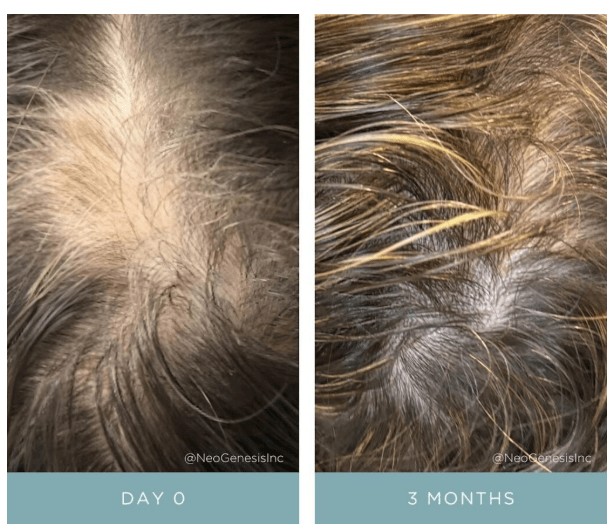
Fu et al (2025) found that hADSC-Exos are more effective in promoting hair follicle development compared to hUCMSC-Exos, and the secretome of ADSCs was more associated with growth processes such as nucleosome function than was the UCMSC secretome (Fu et al, 2025).

Mechanisms Of Action of NeoGenesis Hair Thickening Serum

Topical application of Hair Thickening Serum (HTS) promotes hair growth by two key means: Providing, 1. Skin and hair follicle endogenous molecules from skin and hair follicle stem cells (Adipose mesenchymal stem cells, fibroblasts, and dermal papillae) that drive and maintain the transition from telogen to anagen, and 2. Botanical ingredients normally derived from healthy diets that support hair growth.

Simple topical application of NeoGenesis Hair Thickening Serum, b.i.d., twice daily.

Let’s look at the hair growth cycle, and some of the many factors affecting hair growth. I’ll then explain some the mechanisms by which HTS drives the hair follicle to the anagen phase.

Figure 1. Schematic of the hair growth cycle and the factors that may influence a transition from anagen to telogen vs. telogen to anagen phase. From Natarelli et al, 2023.

HTS Mechanisms of Action in the Hair Growth Cycle

HTS’ mechanisms of action at the hair follicle are many. Here I consider a simplified summary of some of the pathways that the stem cell released molecules and botanical ingredients activate or inhibit to drive and maintain the follicle’s transition to the anagen phase.

Transition from Anagen to Telogen

Inflammation – An immunoprivileged state in the follicle is needed to drive anagen, and inflammation transitions the follicle to telogen instead (Bertolini et al, 2020). HTS reduces inflammation in the innate and adaptive immune systems by using the secretome from adipose mesenchymal stem cells – both the exosomal fraction and soluble fractions that act synergistically to optimally reduce inflammation (González-Cubero et al, 2022; Mitchell et al, 2019)

Hormone – ADSC secretome inhibits negative effects of DHT on hair growth (Tang et al, 2023; Fu et al, 2025).

Poor Nutrition – HTS contains nutrients to support hair growth. Larix Europaea Wood Extract, containing Dihydroquercetin-glucoside (polyphenol), EGCG (polyphenol catechin), glycine, zinc, Camellia Sinensis Leaf Extract, Santalum Acuminatum Fruit Extract, Citrus Glauca Fruit Extract, Acacia Victoriae Fruit Extract, Trifolium Pratense (Clover) Flower Extract (providing an abundance of polyphenols and antioxidants).

Stress – ADSC secretome mitigates immunological disturbances affecting the hair follicle (HF) and contributing to hair loss. ADSCs are able to suppress lymphocyte proliferation and, inhibit complement activation and dendritic cell differentiation from monocytes and therefore are considered natural immunosuppressants (Salhab et al, 2022).

Transition from Telogen to Anagen

Blood Flow – Secretome of ADSCs promotes angiogenesis and increased blood flow to follicles (Silveira et al, 2022; Zhu et al, 2020)

Direct stimulation of Hair Growth – Exosomes from dermal papillae cells drive hair follicle stem cell proliferation to rebuild hair follicle (Li et al, 2023), while fibroblasts provide many building-block proteins need to reconstruct the follicle architecture as it transitions from telogen to anagen (Suh et al, 2023).

Increased Local Growth factors – Fibroblasts (Lin et al, 2015), ADSCs (Won et al, 2017), and dermal papillae (HU et al, 2020) secretome all provide necessary growth factors to induce transition to anagen

References

Bertolini M et al (2020) Hair follicle immune privilege and its collapse in alopecia areata. Exp Dermatol. 29: 703–725.

Fu Y, Han YT, Xie JL, Liu RQ, Zhao B, Zhang XL, Zhang J, Zhang J. Mesenchymal stem cell exosomes enhance the development of hair follicle to ameliorate androgenetic alopecia. World J Stem Cells 2025; 17(3): 102088

Fu Y, Han YT, Xie JL, Liu RQ, Zhao B, Zhang XL, Zhang J, Zhang J. Mesenchymal stem cell exosomes enhance the development of hair follicle to ameliorate androgenetic alopecia. World J Stem Cells 2025; 17(3): 102088 [PMID: 40160691 DOI: 10.4252/wjsc.v17.i3.102088]

González-Cubero, E et al (2022) María L. González-Fernández, Elias R. Olivera, Vega Villar-Suárez,Extracellular vesicle and soluble fractions of adipose tissue-derived mesenchymal stem cells secretome induce inflammatory cytokines modulation in an in vitro model of discogenic pain,The Spine Journal,Volume 22, Issue 7,2022, Pages 1222-1234

Li J, Zhao B, Yao S, Dai Y, Zhang X, Yang N, Bao Z, Cai J, Chen Y, Wu X. Dermal PapillaCell-Derived Exosomes Regulate Hair Follicle Stem Cell Proliferation via LEF1. Int J Mol Sci. 2023 Feb 16;24(4):3961.

Lin WH, Xiang LJ, Shi HX, Zhang J, Jiang LP, Cai PT, Lin ZL, Lin BB, Huang Y, Zhang HL, Fu XB, Guo DJ, Li XK, Wang XJ, Xiao J. Fibroblast growth factors stimulate hair growth through β-catenin and Shh expression in C57BL/6 mice. Biomed Res Int. 2015;2015:730139.

Mitchell R et al (2019) Secretome of adipose-derived mesenchymal stem cells promotes skeletal muscle regeneration through synergistic action of extracellular vesicle cargo and soluble proteins. Stem Cell Res Ther. 10(1):116.

Natarelli N, Gahoonia N, Sivamani RK (2023) Integrative and Mechanistic Approach to the Hair Growth Cycle and Hair Loss. J Clin Med. 2023 Jan 23;12(3):893.

Salhab O, Khayat L, Alaaeddine N (2022) Stem cell secretome as a mechanism for restoring hair loss due to stress, particularly alopecia areata: narrative review. J Biomed Sci. 2022 Oct 5;29(1):77.

Shiqi Hu et al. (2020) Dermal exosomes containing miR-218-5p promote hair regeneration by regulating β-catenin signaling.Sci. Adv.6,eaba1685(2020).

Silveira BM, Ribeiro TO, Freitas RS, Carreira ACO, Gonçalves MS, Sogayar M, et al. (2022) Secretome from human adipose-derived mesenchymal stem cells promotes blood vessel formation and pericyte coverage in experimental skin repair. PLoS ONE 17(12): e0277863.

Suh SB, Ahn KJ, Kim EJ, Suh JY, Cho SB. (2023) Proteomic Identification and Quantification of Secretory Proteins in Human Dermal Fibroblast-Conditioned Medium for Wound Repair and Hair Regeneration. Clin Cosmet Investig Dermatol. 2023;16:1145-1157

Tang, Xin, Cao, Cuixiang, Liang, Yunxiao, Han, Le, Tu, Bin, Yu, Miao, Wan, Miaojian, Adipose-Derived Stem Cell Exosomes Antagonize the Inhibitory Effect of Dihydrotestosterone on Hair Follicle Growth by Activating Wnt/β-Catenin Pathway, Stem Cells International, 2023, 5548112, 20 pages, 2023.

Won CH et al (2017) The Basic Mechanism of Hair Growth Stimulation by Adipose-derived Stem Cells and Their Secretory Factors. Curr Stem Cell Res Ther. 2017;12(7):535-543

Zhu, D., Johnson, T.K., Wang, Y. et al. (2020) Macrophage M2 polarization induced by exosomes from adipose-derived stem cells contributes to the exosomal proangiogenic effect on mouse ischemic hindlimb. Stem Cell Res Ther 11, 162.

DNA Damage Repair: Animals Do It Better Than Plants and Microorganisms

Although plants and microorganisms such as algae possess some of the DNA damage response factors that are present in animal systems, they are missing many of the important regulators, such as the p53 tumor suppressor. The p53 mechanism halts the cell cycle when DNA damage is detected, giving repair machineries time to act. These observations point to the differences in the DNA damage response mechanisms between plants and animals. While DNA repair enzymes from plants may help the skin when topically applied, optimizing the DNA repair mechanisms inherent in animals is the optimal strategy for repairing DNA in animals. Adipose mesenchymal stem cell released molecules (secretome) help to optimize inherent DNA repair mechanisms when topically applied to the skin, including heat shock proteins. More importantly, the molecules help to rebuild the architecture of the skin, which prevents mutations from forming cancerous cells (see my Blog on this topic).

All plant extract contain DNA repair enzymes

If you’re using a product with plant extract, you’re likely using DNA repair enzymes. All, or nearly all, plants contain DNA repair enzymes. Keep in mind, this is important, an intrinsic feature of plant and microorganism DNA repair pathways is that they are not error-free, leading to potentially transmissible mutational alterations. The error-prone nature of some DNA repair mechanisms, however, increases the genetic diversity and variability of the populations, thus contributing to the evolution of plant genomes. In other words, despite the recent hype about DNA repair enzymes in plants/microorganisms, they don’t work well and are “error prone.”

Animals have more robust DNA reair enzymes than do plants

This is not true in animals. They are not error prone. Because animals must better protect their DNA than do plants to prevent and repair mutations that are harmeful, potentially lethal. This is why stem cells in the skin faciltate DNA repair as only animals can do.

Stem cells in the skin have the most robust DNA repair mechanisms

As stated in a presitigious scientific journal, Molecular Cell, ” adult stem cells are endowed with a superior capacity to prevent the accumulation of genetic lesions, repair them, or avoid their propagation to daughter cells, which would be particularly detrimental to the whole organism.” Further stated, “SCs [stem cells] count upon robust antioxidant defenses (which limit genotoxicity) and a superior DDR (to repair unavoidable damage). These mechanisms are in place to protect cellular homeostasis.” 

DNA repair mechanisms: it’s complicated and very efficient if enabled

DNA double strand breaks (DSBs) are a serious threat to genome stability and the erroneous repair of DNA may lead to chromosomal rearrangements with potentially lethal consequences, including cancer, for an organism. The response to DSBs elicits a highly complex and organized cellular program, called the DNA damage response (DDR), setting in motion processes that mitigate the adverse effects of DNA damage and facilitate DNA repair. Broken DNA is usually repaired by two mechanistically distinct pathways: homologous recombination (HR) and non-homologous end joining (NHEJ). HR is a complex, multistep process that allows large sections of DNA to move from one chromosome to another. NHEJ is a DNA repair mechanism that fuses broken DNA ends together without the need for a homologous template While HR uses a homologous DNA strand as a template for error-free repair, NHEJ is inherently error-prone and does not rely on sequence homology. The preferred mode of repair and cellular consequences of DDR varies between organisms and is also dependant on cell type and cell cycle context. For example, while HR is the preferred mode of repair in many unicellular organisms such as budding and fission yeast, NHEJ is the prevalent pathway in plants and animals.

Plants versus animals, digging deeper into the mechanisms

However, in many aspects, plants respond differently to DNA insults than do animals. The constant risk of tumor formation in animals has led to evolution of DDR that assures precise genome maintenance, often resulting in apoptotic death of significantly damaged cells. The lack of such a strong selective constraint presumably permitted evolution of a less potent DDR in plants, making plants more prone to genome damage. Furthermore, plant cells are exposed to high levels of genotoxic stress resulting from long-term exposure to solar ultraviolet (UV) irradiation, photosynthesis and extended periods of desiccation. Thus, some features of plant DDR and DSB repair may deviate from models primarily established from studies in yeasts and mammals.

DNA repair in animals is even more complicated than that described by the two major pathways. Five DNA repair mechanisms are usually distinguished: (a) direct DNA damage reversal, (b) BER, (c) mismatch repair (MMR), (d) nucleotide excision repair (NER), and (e) homologous recombination (HR) and non-homologous end joining (NHEJ). DNA repair pathways were originally restricted to the nuclear compartment. Ample evidence indicates that mitochondria possess a number of DNA repair factors and mechanisms shared with the nuclear processes.

From: Sottile and Nadin (2018). Sources of DNA damage and repair mechanisms. Endogenous and exogenous agents constantly impact on DNA. They may cause many different forms of DNA damage. The scheme shows the five major DNA repair mechanisms operating in the nucleus of mammalian cells capable of removing a wide range of DNA lesions: direct damage reversal, base excision repair (BER), nucleotide excision repair (NER), mismatch repair (MMR), homologous recombination (HR), and non-homologous end joining (NHEJ). The BER system may also be found in the mitochondria. ROS reactive oxygen species, IR ionizing radiation, TOPOII topoisomerase II

Not included in the above summary is a new form of DNA repair in animals, called neucleophagy. This was just reported in October 2024 by a group of scientists at Oxford. Found to be evolutionarily conserved and clinically relevant, we’ll know more baout this mechanism in the coming years. Its mediated by TEX264, an intrinsically disordered protein, and as such, I suspect this will be an important and widespread DNA repair mechanism, possibly involving adult stem cells and their secretome – stay tuned.

How adult adipose mesenchymal stem cell scretome faciltates DNA repair

These complicated processes are faciltated by a number of molecules, including proteins, such as heat shock proteins, released by adipose mesenchymal stem cells (ADSCs). Another example, ADSCs release sirtuins, which are involved in DNA repair. The sirtuins work both as protein activators and chromatin-structure-modifying enzymes. Deacetylation carried by sirtuins represents a basic epigenetic mechanism. Histone modifications including deacetylation and poly-(ADP)-ribosylation compromise an essential part of physiological ageing processes that are involved in the pathogenesis of ageing-related diseases. Stem cells are also known to produce 5-hydroxymethylcytosine binding, embryonic stem-cell-specific (HMCES) protein functions as an intermediate in DNA interstrand cross-link repair, part of BER. Antioxidants from ADSCs are also important for DNA repair. They include: Superoxide dismutase (SOD): This enzyme converts superoxide radicals (O2-) into hydrogen peroxide (H2O2), which is then further broken down by other enzymes like catalase. Catalase (CAT): Catalase directly breaks down hydrogen peroxide (H2O2) into water and oxygen, preventing further oxidative damage to cellular components including DNA.  Peroxiredoxins (Prxs): This family of enzymes also plays a significant role in scavenging reactive oxygen species, particularly in the nucleus where DNA is located, and can directly contribute to DNA repair mechanisms.

The secretome from ADSCs was tested for skin repair following irradiation, where DNA and protein damage is a key component in the radiation dermatitis. Working with Dr, Michael Traub, N.D., NeoGeneis found that simple topical application of ADSC secretome in its Recovery product significantly reduced radiation dermatitis. The Recovery works through the many types of molecules found in the ADSC (and fibroblast) secretome that enable the skin’s robust DNA repair mechanisms to work optimally.

Beyond the hype of plant/microorganism DNA repair enzymes

You can see that using the secretome from ADSCs is much more powerful than using a plant or microorganism extract to repair DNA. While DNA repair enzymes from plants and microorganisms have been found to reduce cyclobutane pyrimidine dimers, and presumably repair DNA in humans when compared to doing nothing, the “DNA repair” products on the market contain many other ingredients that are at least partially responsible for their efficacy. Those other ingredients include antioxidants and sunblocks. If you want a great topical product to repair DNA in your skin cells, use NeoGenesis Recovery that is full of the molecules released from ADSCs, including those molecules in exosomes and those molecules in the soluble fraction.

“Exciting Exosomes in Aesthetic Dermatology” – What Zoe Diana Draelos, M.D. Doesn’t Seem to Understand

In a piece published in the Dermatology Times, Zoe Draelos, M.D. misinforms the dermatological community about exosomes.

Exosomes are an exciting technology, and the complications of this technology are many. I’ve been publishing about exosomes for many years, and if you’d like to read a deep dive into exosomes, you can read my free Elsevier-published review chapter on exosomes that I wrote back in 2016. Named, “Exosomes: smart nanospheres for drug delivery naturally produced by stem cells,” the chapter is available free on Research Gate. You can also read my recent blog on exosomes, and in another blog read about some of the companies bringing sub-optimal exosomes to the market. As I described in my 2013 paper, “Stem Cell Therapy Without the Cells,” using a reductionist strategy where only some of the molecules are used, instead of all the molecules, is a suboptimal strategy. Using only exosomes is reductionistic and suboptimal. My blog, chapter, and papers, explains what Zoe Draelos doesn’t understand about exosomes. Use the secretome (all of which is released by the cell), not just the exosomes. A number of studies have found that exosomes don’t have the same efficacy as does the complete secretome (the natural secretome that contains both the exosome faction and the soluble fraction), including for actions such as immune modulation and regenerative capacity.

I’ll keep it simple here in this blog, and refer to one part of the article by Zoe Draelos. I’ll focus on the following paragraph from her article: “Exosomes for aesthetic use are derived from adult or mesenchymal stem cells. These cells can be harvested from umbilical cord mesenchymal stem cells or adipose-derived stem cells. The exosomes are isolated by differential centrifugation from culture media. The culture media is first centrifuged to remove higher mass contaminants. The centrifugation then occurs at higher and higher speeds until the exosomes aggregate as a pellet in the bottom of the centrifugation tube. These purified exosomes can then be placed into cosmetic formulations.”

While some companies do use damaging techniques to process exosomes, for example ultracentrifugation of the cellular culture media to isolate exosomes, and then lyophilization (freeze-drying) the exosomes to preserve them, some companies, such as my own, Neogenesis Inc, use fresh exosomes that haven’t been damaged by ultracentrifugation and lyophilization processes. Ultracentrifugation and lyophilization are used for the convenience of the companies, allowing the exosomes to be easily stored and easily shipped as a small dehydrated, frozen pellet. Scientists have been isolating exosomes for years. The process is challenging. To better understand exosomes, scientists need to isolate them, but they’re hard to isolate because other molecules, particularly proteins not in the exosome, co-isolate with the exosomes. And the processes used for isolation are damaging. For therapeutic purposes, isolation of exosomes is unwarranted – if you want an optimal product.

Isolation of exosomes is unwarranted for three major reasons: 1. as I discussed, the process damages exosomes rendering damaged proteins on the inside of the exosome as well as those tethered to the outside, and 2. the highly functional proteins and polysaccharides attached to the outside of the exosome can by stripped away – the exosome is denuded, and 3. cells release many beneficial molecules that are not contained in or on the exosomes. When cells release molecules, there is an exosomal fraction and a soluble fraction. The two fractions work together synergistically, and excluding one or the other yields a suboptimal product. In other words, using just the exosomes instead of the exosomes plus the soluble fraction (the molecules secreted by the cell but not contained in the exosomes) yields a suboptimal product.

Exosomal cargo is protected from enzymatic, pH, and heat degradation given its encapsulation within the lipid bilayer of exosomes. Exosomal proteins have been found to maintain their native conformation and functionality for long periods of time, where, for example, exosomal phosphoproteins were stable over a storage period of at least 5 years (Chen et al, 2017). Exosome contain heat shock proteins, for example, that repair proteins and may finish the folding of proteins within the exosome (Maguire, 2016).

Exosomes are complicated and we still have much to learn. But what we have learned is that fresh, unprocessed exosomes work best because they’re undamaged, and when the exosomal molecules are combined with the other molecules that are released by the cell but not contained in the exosomes, we have an optimized therapeutic. The unprocessed exosomal fraction in combination with the unprocessed soluble fraction works best.

Skin Longevity: Mesenchymal Stem Cell Released Molecules Contain SIRT1 and Other Molecules Upregulating SIRT1 Expression

The molecules released by adipose mesenchymal stem cells (ADSC) are known to bring skin cells out of senescence, and the mechanism of action is twofold: 1. the molecules released from ADSC contain SIRT1, and 2. the molecules released from ADSC increase SIRT1 expression in target cells. These are two of the many mechanisms of action underlying the ability of NeoGenesisS2RM technology to rapidly and sustainably reverse and prevent the signs and symptoms of skin aging.

Longevity is a hot topic in the popular press, and the topic has now hit skincare. This is part of the “scientification” of skincare in the popular press that has arisen over the last few years. The trend has an upside and a downside. Learning about ingredients and how they work in the skin is important. The better informed we are, the better we can take care of our skin. The downside, is that non-scientists, including many practicing dermatologist, who have neither been trained as scientists, nor trained to analyze scientific studies, often proffer erroneous information about skin care in the popular press. For example, in my 2020 publication, in the section called “Example of Misinformation in Skin Care Marketing.” I describe how a practicing dermatologist in Miami makes many mistakes in describing skin care ingredients in her various popular press articles, including articles in a large newspaper.

Also, reading a Bloomberg article on what dermatologist think about anti-aging skin care products, I was once again shown an example of how misinformed are some practicing dermatologist. One dermatologist was saying not to use products that contain antimicrobial preservatives, but when looking at the products she has for sale on her website, guess what – many of the products she sells online contain an antimicrobial preservative. Looking at her blog, she talks a lot about using sunscreen – an important topic. But she misinforms the reader by saying that mineral sunscreens reflect light and UV. That’s incorrect because mineral sunscreens predominantly absorb the UV, not reflect it. I didn’t read anymore because I dislike misinformation – I’ll stick to reading informed articles from professors of dermatology, including scientists and physicians, who inform us based on scientific and clinical evidence.

Let’s look at what’s being said about longevity of the skin in the popular media, and have a brief look at some of the science in the scientific literature (PubMed, peer-reviewed journal articles).

Longevity in Skincare

As Jeannette Neumann in Bloomberg states:

In the same article, Neumann goes on to tell us that:

Key to the new product ate sirtuins. So what are sirtuins and what do they do? Sirtuins are a family of signaling proteins involved in metabolic regulation. They are ancient proteins in animal evolution and appear to possess a highly conserved molecular structure throughout all kingdoms of life. They are everywhere in lifeforms. And guess what the scientific evidence suggests: NeoGenesis not only has SIRT1 activators in our S2RM technology, but we also have the SIRT1 protein itself. We’ve had S2RM on the market for over 13 years as a topical product. It’s one of the reasons our products work so well.

Here’s a little more on sirtuins and how they’re activated. SIRT1 is a cellular defense protein that ensures survival by controlling metabolism when there is not enough energy supply to cells. SIRT1 is an important molecule in the control of redox states, apoptosis, and a number of life-extending mechanisms. By changing SIRT1 expression, a number of substances and factors can control the level of SIRT1 protein. Naturally occurring molecules that increase SIRT1 expression include, resveratrol, quercetin, fisetin, curcumin, and berberine. SIRT1 protein expression declines as a we age, and SIRT1 expression decreases with age in mice. SIRT1 has been referred to as a longevity-associated protein that could be used as a potential therapeutic target for extending human healthspan, and it is currently under investigation in the battle against cognitive decline, neurodegenerative diseases, and aging. SIRT1 has been reported to negatively regulate the expression of a number of inflammatory senescence-associated secretory phenotype (SASP) factors, including the SASP factor. SIRT1 produces neuronal protection in neurodegenerative disorders and memory impairment, and is crucial for synaptic plasticity and memory retention in neurons. Numerous studies have shown that p53 and p21 have a role in the control of the cell cycle, DNA repair, apoptosis, and other critical biological processes. Cell cycle arrest results from the activation of p53 and p21, which are responsible for replicative and stress-related senescence in cells. SIRT1 acts on p53 by deacetylating it, which negatively regulates p53’s transcriptional activity, essentially suppressing its function as a tumor suppressor and inhibiting apoptosis induced by stress or DNA damage. In other words, SIRT1 “dampens down” the activity of p53 by removing acetyl groups from it. 

Senescent cells release a range of inflammatory proteins, such as SASP, which causes low-grade chronic inflammation and accelerates senescence. Loss of the key anti-aging molecule SIRT1 may be important for accelerating aging. Zhang et al (2023) found that aged mice displayed upregulation of senescence-related signals such p53 and p21 and downregulation of SIRT1 in the hippocampus. These abnormalities were reversed by the molecules released from mesenchymal stem cells (MSCs).

In our skin, fibroblasts are long-lived cells that are subject to much damage over the years. They can become senescent and pro-inflammatory. Studies have found that SIRT1 can protect human fibroblasts from senescence by promoting telomerase reverse transcriptase transcription (Yamashita et al, 2012). Further, Yuan et al (2012) found that SIRT1 improved the senescence of young MSCs during in vitro subculturing. In other words, SIRT1 protects young cells from stressors, such as oxidative stress, and keeps them healthy and from becoming pro-inflammatory senescent cell types.

As you can see from these studies, it’s not just about anti-aging, it’s about promoting longevity in the first place. We do both at NeoGenesis with our S2RM technology.