Why Seed Oils Are Great For Skin Care

Did you know scientists have discovered that topical sunflower seed oil helps to rebuild the stratum corneum and barrier function? Or that topical safflower oil can help to regrow hair and help to close wounds. Did you know that consuming seed oil, full of polyunstaurated fats, is healthier than eating coconut oil, which is 90% saturated fat. Consuming staurated fat leads to heart problems and dysbiosis, among other problems. Did you know that some seed oils, such as high oleic sunflower seed oil, tends to have higher stability than the high linoleic varieties of oils? Or that safflower seed oil contains extremely stable oleosomes? These are the reason why I formulate with these two oils. They provide essential oils (oils the skin needs to aquire to be healthy but are not made by the skin) and they provide numerous benefits to the skin. If the seed oils are extracted carefully, such as expeller pressed, and then formulated in a cold-process, the seed oils are of great benefit to the skin.

Listening to some unknowledgeable and outspoken people on TikTok, YouTube, including some pharmacists, or any of a number of podcasts, the oil extracted from plant seeds is poisoning us. RFK, Jr., who should have stuck with being an environmental lawyer, has jumped on this inane bandwagon. I think his cerebral arteries must be clogged with beef tallow (50% saturated fat) and advanced glycation end products from the fries he eats. Other people, such as the physician Mark Hyman are telling people to eat unhealthy coconut oil instead of seed oils – I guess he’s trying to drum-up business. Coconut oil induces inflammation and metabolic disorders, and a coconut oil- high fat diet has been found to cause dysbiosis associated with an increased risk of colorectal cancer. Further, diets rich in saturated fats, such as coconut oil, may contribute to mitochondrial dysfunction, protein aggregation, and neuronal degeneration. Recent studies (McCright et al, 2025) also find that saturated fats exacerbate asthma and  promote lung myeloid cell inflammasome activation and IL-1β–mediated inflammation in mice and humans.

Instead of pseudoscience from RFK, Jr, try some science from Prof. Dr. Christopher Gardner, Ph.D. on a recent podcast. “It’s so odd that the internet has gone wild demonizing these things [seed oils],” said Dr. Christopher Gardner, Ph.D., a professor of medicine at Stanford University School of Medicine in California and a nutrition scientist at the Stanford Prevention Research Center. “They are not to be feared.” Dr. Gardner says seed oils contain high levels of omega-6 fatty acids, a polyunsaturated fat the body needs but cannot produce itself, so it must come from foods. Polyunsaturated fats help the body reduce bad cholesterol, lowering the risk for heart disease and stroke. The American Heart Association supports the inclusion of omega-6 fatty acids as part of a healthy diet. Studies of linoleic acid, found in seeds, finds the seed oils benefit cardiovascular function.

Seed Oils in Topical Skincare Products

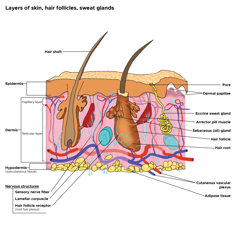
Some seed oils, such as sunflower seed oil, are great for the skin. Sunflower seed oil helps to rebuild skin barrier function. In contrast to sunflower seed oil, topical treatment with olive oil significantly damages the skin barrier, and therefore has the potential to promote the development of, and exacerbate existing, atopic dermatitis.

Sunflower Seed Oil

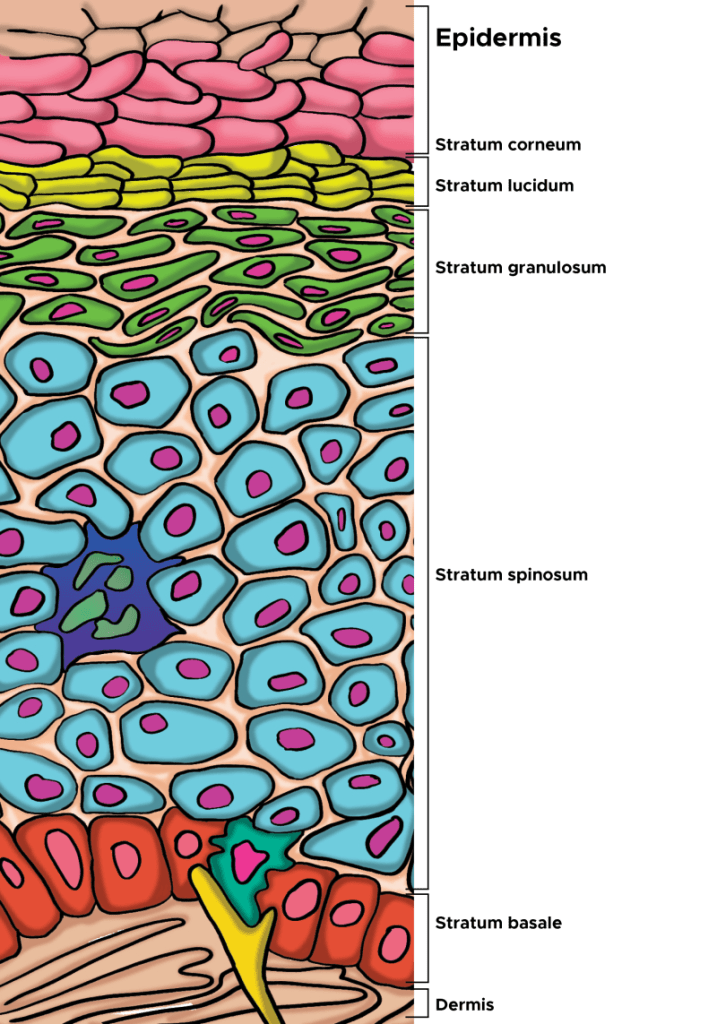
Notice sunflower seed oil (SFSO) is used by dermatologists at UCSF, such as Prof. Dr. Peter Elias, M.D. and his colleagues, in an inexpensive product for building the epidermis and barrier function: https://pmc.ncbi.nlm.nih.gov/articles/PMC9078150/. There are other products that work better, but they are more expensive because of ingredients like oleosomes, Triple Lipid Technology and Natural Mositurizing Factors, things that I’ll explain later. The product developed by Prof. Elias and colleagues features sunflower seed oil, something that is less expensive but provides benefit to barrier function. SFSO activates PPAR-alpha, which has numerous benefits in the epidermis (increased lipid production and lamellar body formation, and differentiation of keratinocyes) that help to rebuild natural barrier function. Sunflower seed oil contains oleosomes if cold-processed and not refined. Oleosomes are specialized plant organelles that protect the oil, and I’ll describe in the next section.

image.png

From Elias et al (2022): The putative mechanisms by which a topical optimised mixture improves permeability barrier homeostasis. In the mixture, petrolatum and lanolin instantly improve the permeability barrier, while glycerol improves barrier (stratum corneum, SC) hydration and accelerates permeability barrier repair. Linoleic acid in both sunflower oil and borage oils activates peroxisome proliferator-activated receptors (PPAR), resulting in increased production of lipids and antimicrobial peptides, and stimulation of lamellar body secretion and membrane maturation. Consequently, both permeability barrier and antimicrobial barriers are improved.

Safflower Seed Oil and its Oleosomes

I’ll also described the benefits of safflower seed oil (SSO), and the oleosomes contained within. SSO has antioxidant and antimicrobial activity, helping to heal wounds. The potential uses of SSO for skin care are under intense investigation given what is already known of this oil. For example, recent results indicate that SSO and its active compound acacetin can prevent UVB-induced MMP-1 expression, which leads to skin photoaging.

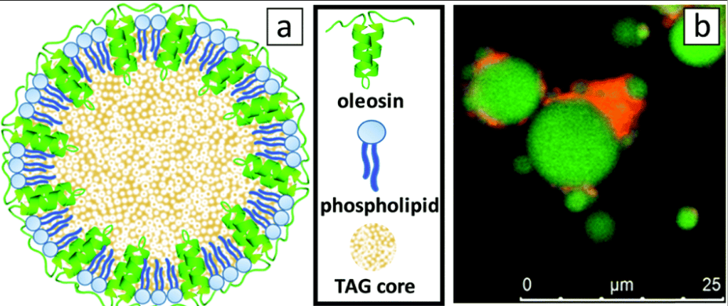
From  Karefyllakis et al (2019): 2D-model of an oleosome with an emphasis on the configuration of the membrane, composed from a monolayer of phospholipids with the hydrophobic oleosin proteins anchored in the triacylglyceride (TAG) core (sizes not to scale), and (b) CLSM (confocal laser scanning microcope) image of SFO stained by Nile Blue showing lipids in green and proteins in red.

Safflower oleosomes are of interest in part because the plant seeds uniquely contain a large amount of oil bodies (oleosomes) and low water content, resulting in decreased hydrolytic properties and lower protein degradation. Within the oil, tocopherol or vitamin E occurs naturally. Looking at the evolution of plant seeds, the primary function of oleosomes is to safely store the seed energy source in the form of triacyglycerols (vegetable oil) during dormancy of the seed and then use it during germination, and to protect the seeds against environmental stressors. To achieve this, a sophisticated structure has evolved where oleosomes are equipped with a membrane that covers the triacylglycerols (TAGs) core, imparting physical and chemical stability. The membrane acts as a shield, preventing the oxidation of the TAGs. The membrane is dilatable and consists of a continuous monolayer of phospholipids that contains a number of proteins, mostly oleosins, embedded in the membrane. Triacylglycerols, also known as triglycerides, are a type of lipid composed of a glycerol molecule bonded to three fatty acids. They are the main constituents of body fat in animals and vegetable fats. Triacylglycerols serve as a crucial energy reserve, storing more than six times the energy of glycogen per gram. The seeds need this energy to germinate.

The oleosomes we use at NeoGenesis are naturally and sustainably extracted from the seeds of safflower (Carthamus tinctorius). The herbaceous non-GMO plant is cultivated and harvested in California, USA, without the use pesticides and with low water requirements. The manufacturer with whom we work, Sharon, uses a proprietary aqueous extraction cold process, comprising: grinding the seeds with aqueous medium, centrifugation and separating the liquids from the solids by centrifugation again before isolating the oleosomes. This process keeps the oleosomes fully intact, maintaining the micro-oil bodies as they occur naturally, with their full qualities and capabilities. Further, cold processing retains the antioxidants, vitamins and minerals. The result is an oil-in-water (o/w) emulsion with natural properties that provide many skin benefits. Biophysical studies of the oleosome have characaterized the oleosomes’ high physical stability, and have called for bioinspired construction of oil structures capable of protection and delivery of ingredients to the skin, all based on the remarkable evolutionary structure of the oleosome.

Sea Buckthorn Oil (Berries, Pulp, and Seeds)

Sea Buckthorn Seed Oil (SBSO), which typically contains the oil from the berries and pulp too, is another highly prized oil for skin care. One of the key reasons I use it in my topical skin care formulations is the high level, about 50%, of Omega-7 fatty acids. SBSO has been found to aid wound healing and increases telomerase activity, and decrease 3-nitrotyrosine. Inhibiting 3-nitrotyrosine (3-NT) is huge because 3-NT is a stress factor and acts as a neoantigen leading to the production of autoantibodies that can destroy skin tissue.

More than Omega-7, sea buckthorn (Hippophae rhamnoides) is valued for its diverse array of bioactive compounds. Cold-pressed extraction is important for retaining the full spectrum of these compounds. The SBSO is typically derived from various parts of the plant, including the whole berry, pulp, and seed, each yielding unique profiles of fatty acids, vitamins (A, C, and E), phytosterols, flavonoids, and carotenoids. SBSO, in particular, is rich in polyunsaturated fatty acids like linoleic acid (omega-6) and linolenic acid (omega-3), as well as the monounsaturated fatty acid palmitoleic acid (Omega-7). SBSO also contains a small amount of saturated fatty acid palmitic acid, monounsaturated fatty acid oleic acid, and trace amounts of myristic and stearic acids, along with minerals such as selenium, copper, zinc, and silicon. Together, this remarkable array of components in SBSO enhance the epidermis’ skin barrier function, structural integrity, and and anti-inflammatory properties, making SBSO a valuable therapeutic agent for conditions such as skin wounds, atopic dermatitis and psoriasis, or any skin condition with inflammation and compromised barrier function.

Other seed oils have been found to provide benefit too. Raspberry seed oil, for example, improve barrier function as measured by decreases of TEWL in human subjects, whereas the comparator oil, coconut oil, had no effect. Let’s stop denegrating seed oils!

Beneficial Seed Oils for Skin

OilKey ComponentsMain Skin BenefitsSkin Types Best Suited For
Rosehip Seed OilLinoleic acid, α-linolenic acid, provitamin A (retinoids), tocopherolsPromotes skin regeneration, reduces scars and hyperpigmentation, improves elasticityDry, mature, scar-prone
Pomegranate Seed OilPunicic acid (omega-5), polyphenolsStrong anti-inflammatory and antioxidant activity, supports skin repair, calms rednessSensitive, inflamed, aging skin
Black Cumin Seed OilThymoquinone, linoleic acidAntimicrobial, reduces acne inflammation, supports wound healingAcne-prone, oily
Chia Seed Oilα-linolenic acid, phytosterolsImproves hydration barrier, reduces trans-epidermal water loss, calms irritationDry, sensitive, eczema-prone
Hemp Seed OilBalanced omega-3 and omega-6, γ-linolenic acidRegulates oil production, reduces inflammation, strengthens barrierOily, acne-prone, combination
Pumpkin Seed OilZinc, tocopherols, phytosterolsPromotes firmness, antioxidant protection, may help with hormonal skin changesAging, combination
Sea Buckthorn Seed OilPalmitoleic acid, carotenoids, tocopherolsHeals damaged skin, boosts elasticity, protects against oxidative stressDry, sun-damaged
Raspberry Seed OilEllagic acid, tocopherols, polyunsaturated fatsAntioxidant, anti-inflammatory, mild natural UV protectionSensitive, aging, photo-exposed

Summary

Bottom line, if you’re using coconut oil or olive oil on your skin, your not improving epidermal function, you can be degrading it. Switch to products using evidence-based, scientific knowledge to formulate their products – and, yes, seed oils, providing essential oils, can be great for the epidermis and improving barrier function. For example, if you use the NeoGenesis Barrier Renewal Cream, you’ll benefit from safflower oleosomes, combined with our Triple Lipid Technology (free fatty acids, ceramide, cholesterol) and Natural Moisturizing Factors (NMF) to rebuild the stratum corneum and barrier function. No other company features this combination of ingredients to rebuild the epidermis. And for those with moderate to severe barrier dysfunction, our new MB-2 product provides instantaneous barrier function without the use of petrolatum, along with 5 types of symbiotic bacteria important to rebuilding barrier function and inhibiting Staphylococcus aureus overgrowth, commonly found in skin with disrupted barrier function. One last thing, NeoGenesis S2RM, containing adipose mesenchymal stem cell secretome, helps to rebuild barrier function too!

For those with disrupted barrier function in conditions such as eczema, including atopic dermatitis, I recommend Skin Serum, which contains S2RM and glycerol, Barrier Renewal Cream, which contains oleosomes+ Triple Lipid Technology +NMF, followed by MB-2, containing a non-petrolatum occlusive+5 live symbiotic bacteria. There’s no better way to improve epidermal health and barrier function, and seed oils are part of the rebuild!.

Skin Pores – Reducing Their Size

Many endogenous and exogenous factors are known to cause enlarged pilosebaceous pores. Such factors include sex, ageing, diet, chronic ultraviolet light exposure, comedogenic xenobiotics, acne, genetic and epigenetic predisposition, and seborrhoea. Most of these causative factors of enlarged pores, being exogenous and controlled by enironmental factors, means you can do something about it. There are procedures and topical products you can use to reduce pore size.

From: Yousef et al (2024)

Although the pathogenesis of enlarged facial pores is still not fully understood, three factors are thought to be key to the pathology: 1) high sebum production, 2) decreased skin elasticity around pores, and 3) increased hair follicle volume. Other factors, including chronic recurrent acne, diet, sex hormones, and skin care regimens, such as inappropriate use of cosmetics, modern Western diets, washing habits, and sun exposure, also affect pore enlargement. Many of these factors will affect the epigenetics of the skin and therefore the skin’s health and potentially pore size. Epigenetics are regulated by your environment, so there is much you can do to reduce enlarged pores.

Causes of Large Pore Size

In cross-sectioned images of conspicuous, enlarged pores, a strongly undulated epidermal–dermal junction was commonly observed around a pore’s opening. Areas with this feature correlated well to the areas with larger hollows and an uneven skin tone. (Sugata et al, 2007).

Recent clinical studies have confirmed the cause of facial pore size to be multifactorial. A positive correlation of pore size and number with sebum output level has been confirmed by several studies (Roh et al, 2006Kim et al, 2013). Enlarged pores increase with age, up to 40 years, and then stabilize or only slightly increase (Jung et al, 2018). Another significant correlation was detected between skin elasticity and pore number in two independent studies suggesting that the structure of dermis could be involved in pore widening (Kim et al, 2013; Hameed et al, 2019). Other observations found pore counts were related to wrinkle severity; and the loss of Microfibril-associated glycoprotein-1 in the hair follicle/pore area with aging and photo-exposure, indicating a lack of matrix support in the dermis (Zheng et al, 2013; Jung et al, 2018).

Both epidermal and dermal structual impairments have been identified as a cause of large pores. Microscopic imaging of pores revealed inner structural changes affecting skin, including a lower density of collagen in the deeper dermis, a thicker stroma and coarser collagen fibers forming a tubular structure around the follicle, and an irregular basement membrane ultrastructure, all of which may result in an altered distribution of skin tensions (Sugata et al, 2008;  Sugiyama-Nakagiri et al, 2008; Mizukoshi and Takahashi, 2014). These ultrastructural alterations may result from inflammation, and recent data suggest inflammaging, mediated by complement activation (immune system proteins), as one of the possible inflammatory agents in the formation of enlarged facial pores (Qiu et al, 2024). Bacteria, such as Staphlacoccus aureus, infect hair follicles and pores, and the question remains, does the inflammation with this sort of infection enlarge the pore. Defects in epidermal morphology around pores have also been discovered, such as epidermis thickening and acanthosis (thickening of the stratum spinosum layer), likely indicating abnormal and possibly excessive keratinocyte proliferation ( Mizukoshi and Takahashi, 2014).

Procedures to Reuce Pore Size

Procedures, such as Micro-focused ultrasound with visualization (MFU-V), have been found to reduce pore size. MFU-V uses focused ultrasound energy to lift and tighten the skin by delivering heat to specific tissue layers beneath the skin’s surface, stimulating collagen production and causing skin tightening according to The Journal of Clinical and Aesthetic Dermatology. The visualization aspect of the procedure allows practitioners to see the underlying tissue during treatment, ensuring precise targeting and optimal results.

Topical S2RM to Reduce Pore Size It’s Not Just the Exosome, It’s the Secretome

But are procedures needed to reduce pore size? No, the right choice of topical skin care products can significantly reduce pore size too. The secretome from adipose mesenchymal stem cells, something used in the NeoGenesis S2RM technology, significantly reduces pore size. That inflammation inducing the ultrastructual changes causing pores to enlarge can be reduced- reduce the inflammation with ADSC secretome found in the NeoGenesis S2RM technology. Remember, It’s Not Just the Exosome, It’s the Secretome that is optimal for reducing inflammation and regenerating tissue – including the tissue that constructs the pore. Changes of TEWL found that ADSC secretome can faciltate the recovery of the skin barrier function (Zhou et al, 2013), which can be explained by ADSC secretome normalizing the proliferation and migration of human primary keratinocytes as reported by Moon et al (2012). Both the epidermis (Ren et al, 2024) and dermis (Silveira et al, 2022) and hypodermis (An et al, 2021) are regenerated by ADSC secretome, with ADSC secretome containing collagen type IV needed to build the basment membrane, thereby regulating that “undulated epidermal–dermal junction” found to underly increased pore size.

I want to emphasie that inflammaging, inflammation that occurs as we age, is exposome induced. Those who eat well and live in an healthy envionment don’t suffer from inflammaging (Franck et al, 2025). As Franck et al write, “Inflammaging, as measured in this manner in these cohorts, thus appears to be largely a byproduct of industrialized lifestyles, with major variation across environments and populations.” In other words, if you live a healthy lifestyle, chronic inflammation, including inflammaging, is something you won’t suffer. This will reflect in your skin health, and your skin’s pore size.

Summary

Pore size in the skin depends on your envionment, your so-called exposome. Healthy skin is beautiful skin, including beautiful, healthy pores. Eat well to keep the skin healthy with sebum production levels normal and therefore reducing a risk factor for increased pore size. And the right choice of topical skin care products can help keep the skin healthy and pore size normal.

NeoGenesis’ New Vitamin C Product – Vibrant C Serum – Why It’s Different and Better

There are many topical vitamin C products on the market, but I needed to formulate something new because the other products are suboptimal for a number of reasons that I discuss here. Liposomal ascorbic acid is one reason the NeoGenesis vitamin C product, Vibrant C Serum, is better, and our built-in antioxidant cascade system is another.

Primates, including humans, cannot synthesize vitamin C. Most other mammals (except guinea pigs), including our cats and dogs, can produce vitamin C, but primates have lost this ability due to a beneficial genetic mutation. Primates evolved to eat mostly, if not exclusively, plants and therefore obtained all the vitamin C they needed through their diets. Look at those big, strong Gorillas, they eat only plants. We primates evolved to eat plants, loaded with C, all that we needed for optimal health, and so we eventually lost the genetics to make vitamin C. In other words, we mutated, and that pesky DNA sequence for making vitamin C was a waste of energy and therefore to be efficient, evolution of primates dropped the unneeded sequence. Pretty cool how mother nature is so efficient and evolution is such as great designer, doing so without a designer. Thinking teleogically, what she said was, “you eat so much vitamin C, you don’t need to make it anymore.”

Reasons Why the Skin May Not Have Adequate Vitamin C Levels

But that was then, and this is now. People don’t eat so well these days and are stressed-out, both of which can lead to suboptimal levels of vitamin C in the skin. Even if you eat sufficent levels of Vitamin C, the transporters of vitamin C from the blood vessels don’t work well under conditions of chronic inflammation – therefore those with chronic inflammation in the skin may not be receiving adequate levels of dietary vitaimin C required for both dermal and epidermal function. The blood vessels bringing the vitamin C to the skin are no longer transporting it out of the blood into the skin – the process has been decommissoned by inflammation in the skin.

Ascorbic Acid (Vitamin C) Doesn’t Easily Penetrate the Skin

Vitaimin C (VC) has a molecular weight of only 176.12 g/mol, but it is a hydrophillic small molecule and because it interacts with water and not lipids, it dosen’t penetrate the fatty stratum coreum very well. Many companies put high levels of VC in their products, but the VC just lays on the surface of the skin.

High Levels of Vitamin C on the Skin’s Surface Oxidize (DHA an Anti-inflammatory) But Still Provide No Benefit

That 15% VC product you’re using may just sit on the surface of your skin where it will oxidize. Dehydroascorbic acid (DHA) is the oxidized form of VC, and it too has anti-inflammatory benefits just as VC does. If only it would penetrate the skin and provide benefit. Oxidized VC in the DHA form has benefits and cells, such as keratinocytes, readily take up DHA to convert it back to VC, but it needs to penetrate to the cells (keratinocytes) in the skin to give benefit. If the DHA sits on the surface of the skin too long, it can further breakdown to oxalic acid that can irritate the skin.

For those who would like to know, DHA is reduced to ascorbic acid by cytosolic reductases GSH-NADPH-dependent, lipoic acid-NADH-dependent, and thioredoxin reductase. The bottom line is that both AA and DHA are important to the skin, but longer term oxidation of AA and DHA into oxalic acid is likely detrimental.

Liposomes Carry Vitamin C Into the Skin

You’ll notice lecithin as part of the ingredient list on the Vibrant C Serum. Lecithin is part of the ingredient combination involved in making the liposomes that encapsulate the ascorbic acid. The liposomes not only protect the ascorbic acid, but importantly, carry the ascorbic acid through the stratum corneum to the deeper layers of the skin. The liposomes act without disrupting the stratum corneum and epithelial barrier function. This is different from some other Vitamin C skin care products that use alcohol as a penetration enhancer. Alcohol, especially at high concentrations, disrupts the lipid structure of the stratum corneum and reduces barrier function.

For example, some products use 15% ascorbic acid, along with an alcohol called Ethoxydiglycol, a type of ethanol. They use over 15% alcohol in their formula because the ethoxydiglycol is listed before the ascorbic acid on the product’s ingredient list – this alcohol is used as a penetration enhancer and using a high level of this alcohol in the formulation can disrupt the corneum stratum and induce irritation. In other words, this product is disrupting the stratum corneum’s barrier function.

Positive Epigenetic and Cellular Effects of Vitamin C Once It’s Absorbed Into the Skin

When VC penetrates to the keratinocytes, remarkable and beneficial physiological processes occur.  VC increases epidermal thickness by promoting keratinocyte proliferation through the DNA demethylation of proliferation-related genes (Sato et al, 2025). This is an epigenetic effect, where VC helps to remove a methyl group that is attached to the DNA in the keratinocyte, so-called demethylation, and this allows the keratinocyte to proliferate. Part of what happens in life is that some of our DNA accumulates methyl group attachements, something that can “turn-off” the DNA. VC demethylates the DNA and turns it on again, allowing the keratinocyte to once again proliferate. It’s much more complicated than this, but for the keratinocytes this is the basic hypotheisis that scientists have brought forth.

In terms of the epidermis, L-ascorbic acid (vitamin C [VC]) is widely recognized for its antioxidant properties in the skin (Masaki, 2010), enhances collagen synthesis (Kishimoto et al, 2013), alleviates UV-induced damage to the epidermis (Kawashima et al, 2018), and inhibits melanin deposition (Sato et al, 2017). Long-term VC deficiency leads to epidermal atrophy in a mouse model with disrupted VC synthesis capabilities (Sato et al, 2012). VC promotes keratinocyte viability, induced expression of differentiation marker genes, and increased SC barrier lipids in monolayer or organotypic cultures of normal human keratinocytes (Boyce et al, 2002Michalak et al, 2021). Together, these data suggest that VC is critical to epidermal cell proliferation and differentiation.

Collagen and Matrix Formation

Vitamin C is also crucial for the production of collagen, a protein that helps to form your skin’s overall structure and barrier and enhances your skin’s elasticity. Collagen is an important building block of the skin. In addition to stabilising the collagen molecule by hydroxylation, vitamin C also stimulates collagen mRNA production by fibroblasts in the dermis yiedling more collagen and stable collagen. Hydroxylation of collagen is a critical post-translational modification where specific amino acids, proline and lysine residues, are hydroxylated, forming hydroxyproline and hydroxylysine, respectively. This process is vital for collagen’s structural integrity, stability, and proper function, particularly within the extracellular matrix. So, Vitamin C helps to stabilize the collagen that is present, as well as to help produce new collagen.

Skin collagen is a long-lived protein, having a long half-life that is estimated to be approximately 15 years, so some skin collagen can exist for much of your life. In young, healthy skin, the amount of enzymes, such as MMP (proteases) that breaks down collagen is low, and therefore, collagen degrades very slowly. However, as we age and more MMP is present, collagen ages, it starts to degrade and fragment. Unfortunately, the degraded and fragmented collagen cannot be incorporated into new collagen fibers and it accumulates within the extracellular matrix of the dermis. The presence of fragmented collagen remainders in aged dermis inhibits both fibroblast proliferation and type I procollagen synthesis by these cells, further degrading the dermis. 

It’s the supporting matrix, such as collagen and elastin, that gives the skin its tightness, thickness, and firmness, but over the years, it starts to break down. That’s why skin gets saggy and thin. Again, Vitamin C is one factor, a necessary factor, to protect collagen and to produce new collagen.

Using the Optimal Amount of Ascorbic Acid: Too Much Vitamin C Inhibits Elastin

Elastin helps form the dermal matrix and helps give elasticity to the skin. Like collagen, much of the eleastin is formed in the dermis by fibroblasts. Differential effects of ascorbic acid on collagen I and elastin mRNA abundance result from the combined, marked stabilization of collagen mRNA, the lesser stability of elastin mRNA, and the significant repression of elastin gene transcription. At NeoGenesis, we use a topical 5% of ascorbic acid, the natural form of Vitamin C, that has been found by scientists at major university in France to rebuld the skin’s ultratructure and provide clincially visible benefits to the skin, rebuilding collagen but not destroying elastin. Moreover, our stem cell-based S2RM technology stimulates fibroblasts to form collagen and elastin, a perfect complement to our Vitamin C product..

Notice we at NeoGenesis don’t use sodium ascorbate because sodium accululates in the skin and induces inflammation, including in skin conditions such as psoraisis and autoimmune diseases. Skin sodium content has been positively and significantly correlated with classical inflammatory markers such as CRP and white blood cell count. For example, dietary salt loading can result in hypertonic sodium storage in the skin by binding to glycosaminoglycans. Topical sodium can add to the accumulation, so wherever possible, we don’t use sodium molecules in our NeoGenesis formulations.

Antioxidant CascadePrimary and Seconday Antioxidants

You’ll find a number of primary and secondary antioxidants (they work indirectly to prevent oxidation) in the Vibrant C Serum, including: Rose Geranium (Pelargonium spp.) water, Ascorbic Acid, Magnesium Ascorbyl Phosphate, Gluconolactone, Ferulic Acid, Panthenol (a secondary antioxidant), Resveratrol, Sodium Benzoate, Hydrolyzed Rice Protein, Potassium Ascorbyl Tocopheryl Phosphate, Chinese Senna (Cassia Obtusifolia) Seed Extract, Glutathione, Curcumin (encapsulated), Honokiol, Magnolol, Ergothioneine, Soybean (Glycine Soja) Protein, and Superoxide Dismutase.

The antioxidant cascade is a series of reactions where one antioxidant molecule neutralizes a free radical and, in doing so, becomes oxidized itself. This oxidized antioxidant can then be recycled back to its active, reduced state by another antioxidant, and so on, creating a chain reaction that efficiently eliminates harmful free radicals. Antioxidants working in a cascade is where one antioxidant type regenerates another type. For example, vitamin E neutralizes lipid radicals but becomes a radical itself; vitamin C can then restore vitamin E to its active form. Enzymatic antioxidants like SOD, CAT, and GPx (Glutathione peroxidase family of enzymes) handle different reactive species at various cellular locations, creating a layered defense This process helps maintain cellular redox balance and protect against oxidative stress. Thus, different types of antioxidants work together in a cascade process by complementing, supporting, and regenerating each other’s activity, resulting in a more robust and comprehensive defense against oxidative stress than any single antioxidant could provide alone. The pathways in cellular oxidation and antioxidation are complex, involving many pathways and may different types of antioxidants. While vitamin C is important, having the other antioxidants available concurrently is critical to the cellular redox balance, the the dynamic equilibrium within a cell between oxidation and reduction reactions. Both are critical to normal cellular function.

Summary

The antioxidant cascade, involving many antioxidant types, not just Vitamin C, is important for neutralizing free radicals, but also very important in regulating cellular signaling, gene expression, and repair mechanisms. Antioxidants, such as the encapsulated curcumin found in Vibrant C Serum, can modulate key pathways (like NF-κB and MAPK) to reduce inflammation and enhance cell survival and regeneration. Delivering these many antioxidants to the cells in the skin where they can provide benefit requires good formulations where, for example, liposome and encapsulation delivery technologies are used by NeoGenesis for its Vibrant C Serum product.

Why NeoGenesis Uses Sodium Benzoate and Gluconolactone As An Antimicrobial Preservative System

The use of gluconolactone and sodium benzoate together as a preservative system has, 1.  a wide range of global regulatory acceptance, 2. Broad spectrum antimicrobial activity, 3.  ECOCERT/COSMOS-accepted. 4.NATRUE- approved and Soil Association-approved, 5. added moisturization benefit, and 6. anti-inflammatory properties. This safe and efficacious preservative system with skin benefits compares to others such as phenoxyethanol that is toxic and easily penetrates the skin into the blood.

Sodium Benzoate

Cinnamon contains a major compound, cinnamaldehyde, which is converted into cinnamic acid by oxidation. In the liver, this cinnamic acid is β-oxidized to benzoate (Abd El-Mawla et al., 2001) that exists as sodium salt (NaB) or benzoyl-CoA. As a safe metabolite of cinnamon, sodium benzoate (NaB), is a widely-used food preservative and a FDA-approved drug against urea cycle disorders in humans, found to increase the levels of neurotrophic factors [e.g., brain-derived neurotrophic factor (BDNF) and neurotrophin-3 (NT-3)] in the CNS (Jana et al, 2013). So safe is NaB that it is approved as an injectable for certain brain diseases (Misel et al, 2013).

NaB is of medical importance as it is a component of Ucephan, a FDA-approved drug used in the treatment for hepatic metabolic defects associated with hyperammonemia such as urea cycle disorder in children (Leonard and Morris, 2002; Scaglia et al., 2004). It is also widely used as a preservative in broad range of foods and cosmetic products (Nair, 2001). It is non-toxic and can be administered as a solution in drinking water. One study reported that a 2% solution of NaB in drinking water is safe for lifelong treatment in mice without any measurable negative side effects (Toth, 1984). Recent studies have found that NaB is capable of modulating both innate and adaptive immune responses (Brahmachari and Pahan, 2007; Brahmachari et al., 2009; Brahmachari and Pahan, 2010), several studies finding that NaB in switching the balance of Th cell subsets toward anti-inflammatory Th2 and Tregs types (Brahmachari et al, 2007; Rezaei et al, 2016). Inflammatory cytokines found in arthritis were also found to be decreased with NaB using in vitro models (Bemani et al, 2020).

NaB is not only efficacious as an antimicrobial preservative and as an immune modulator, but it is also safe – it does not convert to benzene under the conditions of use as a cosmetic or food preservative as told by some ignorent people I’ve heard talk about the subject. This was a concern back in the 1990s, but was cleared as a problem in the 2000s. Those initial reports from the FDA that small amounts of benzene were in soda drinks has now been found to be in error, as the FDA has said, “the TDS [FDA’s Total Diet Study lab] benzene results appeared to be unreliable.” Benzene formation in the analytic techniques used by the FDA’s TDS lab in Kansas were the culprit, along with contamination (FDA Report, 02/25/2022).

Gluconolactone

In nature, GLA can be found in honey, tofu, cheese, wine, bread, fruit juices, among others, and as an approved food additive

Gluconolactone (GDL) is anti-inflammatory by enhancing in vitro induced (i)Treg differentiation and function, and in imiquimod-induced autoimmunity in mice, treatment with GDL alleviates inflammation by inhibiting TH17 cells (Li et al, 2025).

In patients suffering from cutaneous lupus erythematosus, topical application of a GDL-containing cream controlled skin inflammation and improved the clinical and histologic appearance of the skin lesions within 2 weeks (Li et al, 2025).

GLA exhibits antioxidant and moisturizing effects. It protects elastin fibers from UV-induced degradation (Jarząbek-Perz et al, 2023).

NaB and GLA Compared to Phenoxyethanol

Compared to anti-inflammatory and non-toxic NaB and GLA, phenoxyethanol (PE) is known to be toxic to epithelial cells (Wang et al, 2020). At concentrations equal to and/or less than those dosages approved for human use, PE significantly decreased the signaling activity of the Akt pathway in epithelial cells within 30 min, and induced their atrophy and death within 24 h of exposure. Further, PE is known to penetrate the skin when topically applied, having a dermal resorption rate of about 45% in humans – meaning 45% of PE applied topically travels through the skin into the blood (Eckert et al, 2025).

Summary

The use of gluconolactone and sodium benzoate together as an antimicrobial preservative system for skin care not only provides safe and effective, broad-spectrum effects, the combination also provides substantial skin care benefits, including moisturization, UV protection, and anti-inflammatory effects. Carefully choosing every ingredient we put into our products is one reason why NeoGenesis products are safe and efficacious.

References

Abd El-Mawla AM, Schmidt W, Beerhues L. Cinnamic acid is a precursor of benzoic acids in cell cultures of Hypericum androsaemum L. but not in cell cultures of Centaurium erythraea RAFN. Planta. 2001;212:288–293.

Brahmachari S et al (2007) Sodium Benzoate, a Food Additive and a Metabolite of Cinnamon, Modifies T Cells at Multiple Steps and Inhibits Adoptive Transfer of Experimental Allergic Encephalomyelitis1. J Immunol 1 July 2007; 179 (1): 275–283.

Bemani P et al (2020) In Vitro Effects of Sodium Benzoate on the Expression of T Cells-related Cytokines and Transcription Factors in Adjuvant-induced Arthritis Model. Iran J Allergy Asthma Immunol, May2020; 19(Supple.1):43-54.

Eckert E, Jäger T, Leibold E, Bader M, Göen T, Hiller J. Dermal penetration of 2-phenoxyethanol in humans: in vivo metabolism and toxicokinetics. Arch Toxicol. 2025 Mar;99(3):1095-1103.

Jarząbek-Perz S, Dziedzic M, Rotsztejn H, Kołodziejczak A. Evaluation of the effects of 10% and 30% gluconolactone chemical peel on sebum, pH, and TEWL. J Cosmet Dermatol. 2023; 22: 3305-3312.

Li W et al (2025) Gluconolactone restores immune regulation and alleviates skin inflammation in lupus-prone mice and in patients with cutaneous lupus.Sci. Transl. Med.17,eadp4447(2025).

Jana A, Modi KK, Roy A, Anderson JA, van Breemen RB, Pahan K. Up-regulation of neurotrophic factors by cinnamon and its metabolite sodium benzoate: therapeutic implications for neurodegenerative disorders. J Neuroimmune Pharmacol. 2013 Jun;8(3):739-55.

Misel ML, Gish RG, Patton H, Mendler M. Sodium benzoate for treatment of hepatic encephalopathy. Gastroenterol Hepatol (N Y). 2013 Apr;9(4):219-27

Rezaei N, Amirghofran Z, Nikseresht A, Ashjazade N, Zoghi S, Tahvili S, Kamali-Sarvestani E. In Vitro Effects of Sodium Benzoate on Th1/Th2 Deviation in Patients with Multiple Sclerosis. Immunol Invest. 2016 Oct;45(7):679-91.

Wang J, Yang Liu, Wendy R. Kam, Ying Li, David A. Sullivan, Toxicity of the cosmetic preservatives parabens, phenoxyethanol and chlorphenesin on human meibomian gland epithelial cells, Experimental Eye Research, Volume 196, 2020, 108057,

Synthetic Peptides are the Rage in Skincare – Too Bad Most Don’t Work Well

Scientists have known for decades about the benefits of natual peptides derived from our diets. However, topically applied synthetic peptides are eaily broken down in the skin and have considerable difficulty penetratring the stratum corneum, both of which degrade their bioactivity. Some peptides have a high molecular weight, many are hydrophilic in character, and are highly susceptibility to enzymatic degradation, requiring the application of formulation technologies to improve the stability and the penetration of peptides through the skin. On the other hand, natural peptides produced by the human body have developed protection and functional mechanisms. If you’re using S2RM from NeoGenesis, you’re using these natually produced peptides. Learning lessons from mother nature, a few science-based companies have followed nature and are attempting to develop biomemetic peptides that are “protected and functional peptides.” Too bad most cosmetic companies don’t know about and don’t use these “protected and functional peptides.”

Written in the journal, Future Drug Discovery, “Peptides have traditionally been perceived as poor drug candidates due to unfavorable characteristics mainly regarding their pharmacokinetic behavior, including plasma stability, membrane permeability and circulation half-life” (Christina Lambers, 2022).

Peptides, along with exosomes, are “one of the skincare industry’s favorite ingredients right now,” Vogue reported in December 2024. At a Clinique launch event in early 2024, a physician declared peptides to be “the buzzword of the year.” The fashion of peptides seems to be at its peak hype (Fig.1): “Skincare is in its peptides era,” the beauty and fashion website Hypebae announced last month. As I am often asked about peptides, and because most of the literature for the lay public about peptides is incomplete and just plain wrong, I offer my short intro to peptides here. My blog is “sciencey” because it has to be in order not to present peptides in a manner that is not superficial and presents simplified dross.

.

From Maguire (2016)

What are peptides?

Basically, they’re short strings of amino acids (less than 50 amino acids). By contrast, proteins are long strings of amino acids. Peptides can be naturally produced or synthetically made in a laboratory. Naturally produced peptides are synthesized in the body as large precursor molecules (i.e., preproproteins) and are post-translationally (after the proprotein is made) processed and cleaved by proteases to generate their active peptide product. Synthetic peptides are typically made in the lab using a solid-phase peptide synthesis process where one amino acid at time is added to the string.

Problems with Synthetic Peptides

Synthetic peptides have many issues: off target activation of pathways that are detrimental to human health, poor targeting of beneficial pathways, poor penetration, and rapid degradation. Further, these synthetic peptides typically undergo lyophilization, a harsh freeze-druing process that disrupts their structure and further disrupts their safety and efficacy. Peptide aggregation and consequent loss of function is one of many problems in using synthetic peptides.

Further, the chemical synthesis process used to create peptides may introduce impurities or byproducts that can potentially lead to adverse reactions and toxicity issues. As stated in a journal from the American Chemical Society, “the current state of the art in peptide synthesis [synthetic peptides] involves primarily legacy technologies with use of large amounts of highly hazardous reagents and solvents and little focus on green chemistry and engineering” (Isidro-Llobet et al, 2019). Further, Varnava et al (2019) report that, “To date, the synthesis of peptides is concurrent with the production of enormous amounts of toxic waste.” In other words, the current industrial-scale peptide synthesis methods involve using substantial quantities of hazardous reagents and solvents, some of which may contaminate your topical product, while much of it pollutes the environment.

Synthetic peptides are cheap to make, and can easily be made in large quantities. That’s a plus for the compnies making them. But synthetic peptides can be problematic for therapeutic interactions that require post-translational modifications that are difficult to incorporate synthetically, such as glycosylation, for biological activity. Difficult to create disulfide linkages, important for the functional attributes of peptides and proteins. And peptides lack efficacy in biochemical pathways where the secondary or tertiary structure is critical or in making larger bioactive peptides and proteins. Natural peptides and proteins don’t suffer from these problems.

Microproteins ( you haven’t heard about these because they’re newly disovered) are Different from Peptides

You haven’t heard about these small proteins because they’re newly discovered and very difficult to identify and characterize. Microproteins are typically less than 100 amino acids (AAs) in length, but are different from peptides because they are not cleaved from a proprotein or protein. Until recently, microproteins have evaded detection because traditional genome annotation methods relied on stringent rules to distinguish protein coding RNAs versus non-coding RNAs (ncRNAs) to minimize the discovery of false positives including a minimum ORF length of 300 base pairs (bps). This ad hoc 100-codon threshold was initially selected based on the calculated probability that ORFs over 300 bps are significantly more likely to encode stable proteins. sORF-encoded microproteins have emerged as important new players in cellular biology and physiology, and they continue to be identified at high rates. To be clear, microproteins are polypeptides originating from short open reading frames (sORF) of less than a hundred codons. For a long time, they have been understudied because it is difficult to distinguish coding from non-coding sORFs. In recent years, the number of putatively translated sORFs has been narrowed down from hundred-thousands or millions to various thousands, owing to the advent of ribosome profiling and advances in bioinformatic and proteomic techniques. Therefore, efforts are now being made to include sORFs with robust translation evidence into databases such as GENCODE. The field of microproteins has since steadily grown, though it is still unclear how many functional coding sORFs exist in the human genome, and relatively few microproteins have been characterized to date. 

Microproteins are made in adipose mesenchymal stem cells (ADSCs) and likely released for therapeutic effect by the ADSCs (Bonilauri et al, 2021). Because of the past difficulty in identifying these microproteins, they are just now receiving attention for their therapeutic value. Understand, microproteins likely provide therapeutic value to the ADSC secretome, but our understanding of this is in its infancy.

Natural Peptides Drived from Diet

Lunasin is a 43-amino acid polypeptide originally discovered in soy. Research into the properties of lunasin began in 1996, when researchers at the University of California-Berkeley observed that the peptide arrested mitosis in cancer cells by binding to the cell’s chromatin and breaking the cell apart. The name of the peptide was chosen from the Tagalog word lunas, which means “cure”. Since its discovery, scientists have identified lunasin as the key to many of soy’s documented health benefits and it has been studied for various benefits, including cancer prevention, cholesterol management, anti-inflammation, skin health, and anti-aging. Lunasin exhibits different biological and chemopreventive properties including anti-inflammatory, anticarcinogenic, antioxidant and immune-modulating properties, anti-atherosclerosis, and osteoclastogenesis inhibition potential. Sounds great, right? But there’s a catch. Lunasin as part of a whole soy diet confers health benefits and is bioavailble, measured in human blood, but isolated lunasin has not shown such beneficial results. Isolated peptides don’t work as well as those peptides in their natural state. This is but one of many examples of natural peptides that are derived from a healthy diet providing benefit in their natural state, but when isolated, not so much happens.

Conjugated Peptides

The the human body, peptide conjugation is a crucial process involving the attachment of chemical entities to peptides to enhance their safety, efficacy, and pentration properties. These modifications, called post-translational modifications, can significantly improve characteristics like stability, targeting, and half-life of the peptide within the body, including the skin. Without peptide conjugation, the peptide is bare, subject to rapid degradation, and hydrophilic (meaning loves water and hates fat) so that it is repelled by the fatty nature of the stratum corneum. These are the problems with most synthetic peptides – they’re not conjugated. They’re not biomemetic but are cheap and can be easily hyped during the peak of the peptide-hype curve, i.e. peak of inflated expectation. However, synthetically conjugated peptides often don’t work well either.

For example, Palmitoyl Pentapeptide-4 is a conjugated peptide (pal-KTTKS). The molecular weight of Palmitoyl Pentapeptide-4 is 802.068 g/mol, larger than the “500 Dalton rule.” It consists of a pentapeptide (a chain of five amino acids, KTTKS) linked to a palmitoyl group, which is a fatty acid. This conjugation somewhat enhances its ability to penetrate the skin and makes it more effective as an anti-aging ingredient. However, Choi et al (2014) found that although pal-KTTKS was more stable than KTTKS, in dermal skin extract, 9.7% of pal-KTTKS remained after 120 min and 11.2% of pal-KTTKS remained at 60 min in the skin homogenate. In the epidermal skin extract, the concentration of pal-KTTKS throughout the 120-min incubation period was almost similar to its initial concentration. Lower amounts of proteolytic enzymes in the epidermal skin extract than in the dermal skin extract and the skin homogenate may account for pal-KTTKS lasting longer in the epidermal skin extract. 

Natural Proteins and Peptides Penetrate the Skin Better than Synthetic Peptides

Palmitoyl Pentapeptide-4 (PP-4), a conjugated peptide. Choi et al (2014) found only 11% of the peptide remains after 60 minutes in a homogenate of dermis – it’s broken down to be ineffective. Further, in skin permeation experiments, no detectable levels of KTTKS and pal-KTTKS (PP-4) were observed in the skin over a period of 48 h – the peptide dosen’t penetrate the skin.

Contrast this to the stem cell released molecules (Secretome) of ADSCs that do penentrate the skin and activate collagen production and a number of other beneficial physiological pathways. The secretome from ADSCs is loaded with proteins, peptides, microproteins, microRNA (not mRNA or DNA) and other beneficiary molecules. They work effectively, as mother nature intended, doing so collectively so that the mutlitude of molecules working togther, a systems therapeutic, exhibit synergistic, beneficial effects.

Summary

Synthetic peptides are all the rage now in skincare. Too bad most of them offer much hype and little or no benefit. There are some newer, more sophisticated conjugated peptides that I’m testing to determine whether they exhibit better efficacy than the current conjugated peptides. Stay tuned.

AnteAge, Founded by a Physician Whose Medical License Was Revoked and is Now Owned by Private Equity, Invents Fake Technology

John Sanderson, whose medical license was revoked for sexual misconduct and repeated negligence, has sold his company, AnteAge, to a private equity company. Now that the PE company has taken over, their marketing people have invented a new word, “Biosome.” for what is called by scientists, a “liposome.”

How do we know the Private Equity guys who own AnteAge are using fake technology? Look at the staement from their website: “Currently the AnteAGE MD bottles do not mention the new Biosome ingredient. As part of our commitment to sustainability, we have chosen to utilize our existing inner packaging rather than generating waste unnecessarily. Please rest assured that your product does in fact have Biosomes included. Please reference the ingredient listing here. Reach out with questions or refer to anteage.com.”

Here’s the statment from their web:

How Do We Know They’re Faking It

If there were actually something new in the bottle, they would have to, by law, relable the product. In other words, because they have only changed their marketing hype, and not the product’s technology, they don’t need to make any changes to the bottle labeling – specifically the bottle’s listing of ingredients. The label and the ingredients remain the same and the only thing changing is what they call the product. There’s no validation in peer-reviewed literature or patent filings confirming a unique mechanism under the name “Biosome.” Rather, it’s just marketing hype, or as some would call it, BS.

So what’s in the bottle? Liposomes. Look at the ingredients on the bottles with the new, fake technology. The list includes “Phosphatidylcholine.” Guess what are made with Phosphatidylcholine. Answer – liposomes! So now AnteAge is calling liposome, you guessed it, Biosomes. This is Private Equity at work. Say anything, do anything, for profit.

Here’s the bottle saying “Biosomes”:

And here’s the bottle’s ingredient list for the “Serum”:

Serum Ingredients:
Water (Aqua), Human Bone Marrow Stem Cell Conditioned Media, Cetyl Ethylhexanoate, Niacinamide, Dimethyl Isosorbide, Polyacrylate-13, Glycerin, Hydrolyzed Myrtus Communis Leaf Extract, Butylene Glycol,
Carbomer, Polysorbate 20, Palmitoyl Tripeptide-1, Palmitoyl, Tetrapeptide-7, Polyisobutene, Benzyl Alcohol, Salicylic Acid, Sorbic Acid, Sorbitan Isostearate, Carnosine, Ilex Paraguariensis Leaf Extract, Maltodextrin,
Disodium EDTA, DOTAP, DSPC, DSPE, DSPE PEG, Sodium Chloride, Disodium Phosphate, Potassium Phosphate, Potassium Chloride, Phosphatidylcholine, Phosphatidylserine, Sphingomyelin, Cholesterol, Mannitol,
Trehalose, sh-Oligopeptide-33, sh-Polypeptide-58, sh-Polypeptide-5, sh-Polypeptide-2, sh-Polypeptide-67, sh-Polypeptide-66, sh-Polypeptide-10, sh-Polypeptide-3, sh-Polypeptide-62, sh-Polypeptide-14,
sh-Oligopeptide-2

Bottome line. Private equity is ruining many things and now they’re lying to the public about skin care ingredients.

If you’d like to read about the science of exosomes and liposomes, you can read my 30 page academic book chapter, peer-reviewed, that I published in 2016 with Elsevier, called Exosomes: smart nanospheres for drug delivery naturally produced by stem cells.

Why I Formulate With Chondrus Crispus Extract, and Why It’s Not Comedogenic

Chondrus crispus extract is a polysachharide, which are not comedogenic, and is known for its anti-inflammatory, moisturizing, and wound-healing properties on human skin.

NeoGenesis is a biotech company that has the most advanced skin care products on the market, utilizing, for example, our S2RMstem cell released molecules technology (exosomes, ectosomes, and soluble fraction) that is the most advanced penetration technology in the skin care marketplace. At NeoGenesis we feature science-to-market ingredients that work and are backed by scientific and clinical studies. Chondrus crispus extract is one the science-to-market ingredients used by NeoGenesis. I’ll dig into the science of Chondrus crispus extract (CCE) in the next paragraph, but even a cursory online search of the ingredient gives you an outline of how good this extract is for the skin. Whether it’s SpecialChem, EWG, or Paula’s Choice, scientists reviewing the studies of Chondrus crispus extract all extole its virtues in skin care. Little wonder the ingredient is widely used in skin care products.

Recent studies have found CCE mitigated inflammation and improved scratch-wound healing, and reduce environmental stress. A number of beneficial metabolites can be obtained from algae, including antioxidants, mycosporine-like amino acids, carotenoids, pigments, flavanoids, and polysaccharides. Further, CCE is sustainably sourced unlike a number of competing ingredients.

CCE also contains 15 of the 18 essential elements that make up the human body. This includes calcium, sulfur, magnesium, potassium, vitamin A, and vitamin K. Further, because CCE contains sulfur, it may help to reduce sebum production. CCE also contains omega-3 fatty acids, good for the skin, including acneic skin, whether topical or oral.

You can also read what other scientists and physicians say about the benefits CCE when topically applied to the skin in the popular press here, and here.

Why I Don’t Formulate Products with SLS

Despite the years of research on the ill effects of SLS (sodium lauryl sulfate), I continue to hear that people, including dermatologists, are using products with this ingredient, including shampoos.

If you’ve ever Googled the causes of a skin irritation or damaged hair, you’ve likely seen posts about SLS, or sodium lauryl (or laureth) sulfate, a common ingredient in beauty products, cleansers, shampoos, toothpastes, and cleaning products.

So what does this ingredient do, why is it in everything, and what does the evidence say about how safe it is?

When we use a cleanser or shampoo, the product usually contains a detergent. That detergent is called a surfactant. A surfactant allows the oil and water molecules to bind together – it’s what’s found in soaps and detergents so we can wash our oily faces or dishes with water and remove the grime.

Sodium lauryl sulfate (SLS) is a surfactant, and its efficacy, low cost, abundance and simplicity mean it’s used in a variety of cosmetic, dermatological, and consumer products.

Our skin’s outermost layer, the stratum corneum of the epidermis, is specially designed to keep harmful things out, and this is where a surfactant can cause problems. Using chemicals that weaken this barrier defence mechanism can potentially cause our skin harm.

As the outermost layer of the epidermis, the stratum corneum is the first line of defense for the body, serving an essential role as a protective skin barrier against the external environment. The stratum corneum aids in hydration and water retention, which prevents skin cracking, and is made up of corneocytes, which are anucleated keratinocytes that have reached the final stage of keratinocyte differentiation (From Murphrey et al, 2022).

Some surfactants are more irritating to our skin than others. For something to be harmful, irritating or allergenic, it has to fulfill two criteria. It has to have been found in studies to irritate human skin, and it has to have the ability to penetrate the skin. SLS does both. It penetrates the stratum corneum and induces an immune reaction, and degrades the structure of the barrier.

Scientists in Germany tested 1,600 patients for SLS irritancy and found 42% of the patients tested had an irritant reaction. Another study, on seven volunteers over a three and a half month period, found regular contact caused irritation, and the irritation subsided once the skin was no longer exposed to SLS. Another study found the warmer the water used with SLS, the more irritating it will be.

SLS is a well established irritatant and is used as a positive control in dermatological testing. That is, new products being tested to see how irritating they might be to human skin are compared to the known irritant, SLS. If a person is sensitive to SLS, they might find the area that has been in contact is red, dry, scaly, itchy or sore. It’s also important to note there’s no scientific evidence SLS causes cancer, despite what is often posted on the internet. So, it’s probably OK to use SLS in products that are used for household cleaners.

Who should avoid SLS?

Everyone, especially people with a history of sensitive skin, hyperirritable skin and patients suffering from skin conditions such as atopic dermatitis (eczema), rosacea, and psoriasis are best to avoid products containing SLS. If you think it might be SLS causing a skin irritation, stop the use of the product and look for products that don’t contain SLS.

Epithelial Barrier Dysfunction in Noncommunicable and Communicable Diseases

The modern world’s dramatic increase in the number and types of chemicals in which man is exposed, a major part of of someone’s exposome, responsible for about 90% of diseases (not genetics), is causing a dramatic rise in noncommunicable and communicable diseases. Over 350 000 chemicals and mixtures of chemicals have been registered for production and use, up to three times as many as previously estimated, and an underestimate of the true number of chemical types that have been produced and commercialized. As the skin and other epithelial tissues are compromised and exposed to communicable diseases, skin and epithelial transmitted diseases are on the rise. For example, the shingles virus can enter through the skin or the epithelial tissue in our respiratory tract, and having shingles can even lead to increased risk of dementia (2nd Ref). Further, a compromised skin epithelial barrier caused by environmental factors such as mechanical trauma, exposure to exogenous proteases in microorganisms and our food, detergents, and air pollution can activate the innate and adaptive immune systems, inducing keratinocytes to release pro-inflammatory cytokines and chemokines and enhancing the antigen presentation by intradermal Langerhans cells (LCs) and dermal DCs and activating T-cells. In turn, for example, activation of T2 type T-cells leads to IL-4, IL-5, and IL-13 secretion, provoking skin barrier alteration, immune cell infiltration into skin, and itch as observed in atopic dermatitis. 

The first essential step to skin immunity is the epithelial barrier, as infection and resulting inflammation are impossible without first breaching it. Epithelia, coated with a sugary glycocalyx, not only comprise our skin but also the mucosal membranes that line our organs. Their ability to secrete squalene, mucus, lipids, and antimicrobials help protect against pathogen invasion. Additionally, epithelia can prevent inflammation by physically shoving out cells infested with toxins, allergens, antigens, pathogens, or other damage by seamlessly extruding them. This is a strategy employed by not only epithelia, but also our hair does the same as it sheds. Given that chronic inflammation could stem from a defective epithelial barrier, the current approach of treating only the inflammation will only partially mitigate symptoms of a more central problem, ongoing wound healing and disrupted barrier.

Scientists now understand that in patients with allergic disease, regardless of tissue location, the homeostatic balance of the epithelial tissue barrier is skewed toward loss of differentiation, reduced junctional integrity, and impaired innate defense and a hyperactive adaptive (trained immunity) immune system. Importantly, epithelial dysfunction characterized by these traits appears to pre-date a predisposition to immunological responses against a range of antigens or allergens, and development of allergic disease.

From the disease perspective, trained immunity is beneficial, as it improves the host’s defense against subsequent infection from pathogens. However, it can also be detrimental and result in overly active immune responses or chronic inflammation.  Even the innate immune system has some memory, given evidence that components in House Dust Mite extract activate and likely train macrophages to produce high amounts of CCL17, IL-6, and cysteinyl leukotrienes following re-exposure to HDM through the TNF-α and PGE2 pathways. Thus, an activated immune system, one that has memory and is primed to react, can lead to sensitivities that may be triggered by an overabundance of chemicals in the environment, and those sensitivities heightened by a disrupted barrier.

Evidence that epithelial barrier dysfunction explains the growing prevalence and exacerbations of inflammatory diseases such as eczema has grown through many studies performed world-wide. Diseases encompassed by the epithelial barrier theory share common features such as an increased prevalence after the 1960s that cannot be accounted soley by the emergence of improved diagnostic methods. They are indeed increasing in prevalence, i.e. the number of afflictions per 1,000 people.

Eepithelial barrier dysfunction enables the microbiome’s translocation from the skin’s surface to interepithelial and deeper subepithelial areas, doing in combination with allergens, toxins, pathogens, and pollutants. Thereafter, microbial dysbiosis and possible infection, characterized by colonization of opportunistic pathogenic bacteria and loss of the number and biodiversity of commensal bacteria results. Local inflammation, impaired tissue regeneration, and remodeling characterize the skin that suffers from impaired barrier. For example, commensal bacteria on the skin’s surface are important for epidermal lipid synthesis and improve barrier function. The skin’s microbiome is therefore critical to maintaining epidermal barrier function. The infiltration of inflammatory cells and inflammatory cytokines to affected tissues is part of the immune system’s response to erradicate invading bacteria, allergens, toxins, and pollutants away from the deep tissues. As Peter Elias, M.D. has written, “AD [atopic dermatitis] can be considered a disease of primary barrier failure, characterized by both a defective permeability (Proksch et al., 2006, and references therein) and antimicrobial function.” Further, inflammatory cells and inflammatory cytokines that migrate from the skin to other organs may play roles in the exacerbation of various inflammatory diseases in other organs. Thus, inflammation iniated in the skin may contribute to chronic inflammatory diseases in other tissues.

What Dr. Elias has been saying is that the permeability-barrier abnormality in AD is not merely an epiphenomenon but rather the “driver” of disease activity, an “outside–inside view of disease pathogenesis” (Elias and Feingold, 2001). The evidence for this is: (1) the extent of the permeability-barrier abnormality parallels severity of disease phenotype in AD, (2) both clinically uninvolved skin sites and skin cleared of inflammation for as long as 5 years continue to display significant barrier abnormalities, (3) topical artificial barrier therapy comprises effective ancillary therapy, and (4) specific replacement therapy, which targets the prominent lipid abnormalities that account for the barrier abnormality in AD, not only corrects the permeability-barrier abnormality but also comprises effective anti-inflammatory therapy for AD (Figure 1Chamlin et al., 2002). Thus, inflammation in AD may begin with insults from without, i.e. the exposome.

That barrier insult can then activate epithelial cells in the skin, keratinocyes, which are non-professional immune cells, but do possess MHC-II molecules, that present antigens to professional immune cells, such as T-cells. Thus, with disrupted barriier, the keratinocytes can recognize antigens and present them to the immune system, leading to inflammation. More and more, scientists are discovering how epithelial cells are part of the immune system, regardless in which organ they exist. Key here is to protect barrier function in all of our epithelial tissues, including the skin.

So if inflammatory diseases such as eczema and psoriasis are environmentally triggered and lead to barrier dysfunction and resultant inflammation, what can we do?

First, calm the inflammation. It’s destructive and further degrades the epidermal barrier. S2RM technology (in NeoGenesis Recovery) is great for reducing inflammation, doing so in both the innate and adaptive immune systems.

Second, use a topical product that provides the 3 lipids and natural moisturizing factors that are needed to rebuild normal stratum corneum and barrier function. One product to use is NeoGenesis Barrier Renewal Cream (BRC).

Third, use a product that provides instantaneous barrier function and commensal bacteria. The instantaneous barrier allows the BRC to rebuld the natural barrier function over time, and the commensal bacteria help to rebuild the barrier through activation of lipid synthesis by skin cells. The commensal bacteria in Neogenesis MB-2 also help to reduce the Staphylococcus aureus infection often assicated with disrupted barrier function.

So remember, these inflammatory skin conditions are triggered by the environment. Therefore, their treatment and prevention means that if you change your environment, you can prevent or treat these diseases. Part of changing your environment is the careful choice of topical products to reduce inflammation and renormalize the structure and function of your skin.

It’s the Skin’s Architecture, Not So Much its DNA, That Causes Skin Cancer

DNA mutations in normal skin occur at high rates without cancerous growth. But when the skin’s architecture is broken down, those mutations can lead to cancer. Maintaing the skin’s architecture is critical to skin health.

Mutations Are Everywhere, But Cancer Isn’t

Scientists have looked at UV-exposed eyelid skin of middle-aged adults, and found that a square inch of normal, non-cancerous skin was riddled with mutations, many of them considered cancer drivers. The number of mutations in normal skin tissue rivaled the number seen in skin tumors, and exceeded the number of mutations seen in other tumor types, like breast cancer. Such findings once again set researchers’ expectations about how powerfully these mutations could promote cancer. There’s more to cancer than just mutations in our cells.

It’s The Architecture Stupid, Not the DNA

Prof. Dr. Cyrus Ghajar, Ph.D., a scientist at Fred Hutchinson Cancer Center, has noted that cancer-driving mutations are defined using animal studies. After identifying what is thought to be a common cancer-associated mutation in human cancers, researchers introduce the mutations into mice to see if tumors arise. If they do, they’re considered cancer drivers. But when you find these mutations in people in normal tissue, then what does that mean? It’s clearly not a driver.  Mutations, it turns out, needs partners to drive cancer. They need another powerful mutation and an abnormal microenviornment, to induce cells toward cancerous growth.

The mutation-riddled reality of normal skin tissue prompts us to realize that skin has ways of handling mutations and keeping cellular growth normal. As Prof. Dr. Mina Bissell, Ph.D. at Berkeley has taught us, our organs are set up for function, and that function is inextricably linked to chemical envionment of the cells and the architecture into which the cells are embedded. Most cells in an organ are differentiated, meaning they perform a specialized function. And this differentiated state isn’t merely governed by an internal molecular decision-making process within each cell. It’s a collective process, a top-down process, where the architecture dictates function. If a cancer cell wanders into another organ and survives, it falls under the spell of the architecture, the top-down process instructing the cancer cell to renormalize. Dr. Bissell taught us this many years ago. As shes says, “to understand cancer it is important to understand that the phenotype can override the genotype.” Further, “influences such as what you eat, your internal metabolism, inflammation and the sun’s rays” affect your phenotype and hence your genotype. For example, in the aforementioned study of eyelids, the sun is causing mutations, but the phenotype, the cellular chemistry and architecture, has overridden the genotype, the mutated DNA, and the cells are behaving normally without cancerous growth.

Cancer Reverts if Normal Architecture is Restored

Dr. Bissell and team, in a landmark study, found that if they took breast cancer cells and put them back into a normal microenvionment, a normal architecture, then the cancer cells reverted back to normal. Their results demonstrated that the extracellular matrix, i.e the architecture and its inherent chemistry, dictate the phenotype of mammary epithelial cells, and thus in the model system tested, the tissue phenotype was dominant over the cellular genotype.

A Glimpse at the Big Picture of DNA, Cells, Architecture and Downward Causation

In the big picture, what I’m talking about is downward causation. The architecture instructs the pieces what to do. So the cellular structure is instructing what the DNA, all of the DNA, needs to do. That’s downward causation. We inherit downward causation because life derives from the cell. Cells make cells. Put DNA in a dish, it sits there, inert. Put DNA into a cell, it will begin to function, with that function dependent on what cell it is in. The cell, of course, has architecture, and it is the cell’s architecture that sets boundary conditions, instructing the molecules in the cell, including the molecules in the DNA, what they should do. We humans arise from cells, the mother’s egg – and that egg receives architectural signaling from the fathers sperm, which delivers DNA contained in it own architecture, the centriole. In other words, that cellular architecture and that of it’s surroundings, is critical to the cell’s function, to creating life, and whether cells will become cancerous. Along with Dr. Mina Bissell, Prof. Dr. Dennis Nobel, Ph.D., at Oxford, has been a pioneer in this way of thinking.

Sun Exposure Can Damage the Architecture, Not Just DNA

Concerning sun exposure and skin cancer, what happens when UV damages the skin? Is DNA damaged? Yes. But damaged too is the architecture, incuding the constiuent proteins and lipids in the architecture. As Drs. Bissell and Ghajar have taught us, it’s the cells surrounding architecture that will determine whether a cell becomes cancerous. So the UV damage of the proteins and lipids that make the architecture of the skin will be critical to determing whether the skin is cancerous or normal.

What to Do to Protect the Skin’s Architecture

What do you need to do for your skin to be healthy and free from cancer? Normalize the architecture of the skin. How do you do this? 1. First, dose your skin with sunlight in moderation to protect the skin’s architecture. If you’re out for long, wear a sunblock. 2. Eat well. Fruits and vegetables contain many of the nutrients to needed to maintain and regenerate the skin’s architecture. 3. You can also utilyze a skin care routine that maintains and regenerates the skin’s architecture. Using a combination of NeoGenesis Recovery and Barrier Renewal Cream, for example, will help to maintain and regenerate the architecture of the dermis and epidermis. NeoGenesis Recovery will also help to optimize the skin’s natural ability to repair DNA. Also available are retinoid products and antioxidant skin care products that can also help to prevent damage and rebuild the skin’s architecture.